Download
1 / 78
780 likes | 806 Vues
Learn about the essential functions of blood, its components, and cell formation, including details about erythrocytes, anemia, leukocytes, and platelets. Explore blood disorders, human blood groups, and the cardiovascular system.

E N D
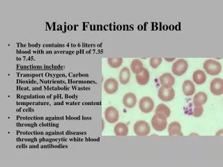
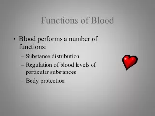
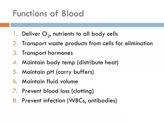
Functions of Blood • Deliver O2, nutrients to all body cells • Transport waste products from cells for elimination • Transport hormones • Maintain body temp (distribute heat) • Maintain pH (carry buffers) • Maintain fluid volume • Prevent blood loss (clotting) • Prevent infection (WBCs, antibodies)
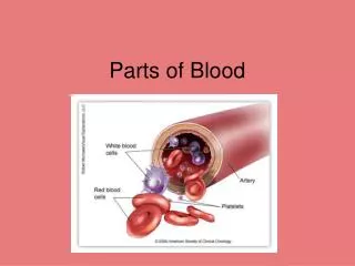
Blood Components • Plasma (55%) • water (90%), ions, proteins, gases, nutrients, wastes, hormones • Cells (45%) • RBCs, WBCs, platelets • Develop from stem cells in bone marrow
Blood Cell Formation • Hematopoiesis: blood cell formation • Occurs in red bone marrow • Skull, pelvis, ribs, sternum, humerus, femur
Erythrocytes • Red blood cells (RBCs) • Transport O2 in blood • Biconcave discs • Anucleate (no nucleus) • Hemoglobin: iron-containing protein, binds to O2 • Life span: 100-120 days
Anemia: decrease in oxygen-carrying ability of blood • Low RBC count or deficient hemoglobin content • Sickle-Cell Disease: abnormal hemoglobin • Genetic disorder • Carriers of 1 allele are resistant to malaria in Africa
Leukocytes • White blood cells (WBCs) • Defend body against infection and tumors • Locate areas of tissue damage by responding to chemicals • Types: neutrophils, eosinophils, basophils, lymphocytes, monocytes
Leukemia: bone marrow becomes cancerous huge numbers of WBCs Treatment: chemotherapy, radiation, stem cell transplant
Platelets • Cell fragments (irregularly-shaped bodies) • Needed for clotting blood
Hemostasis= stoppage of bleeding • Vascular spasm • Constrict damaged blood vessels • Platelet plug forms • Platelets stick and bind to damaged site • Release chemicals to attract more platelets • Coagulation • Blood clotting • Fibrin threads forms mesh that traps RBCs Time: blood clot normally forms within 3-6 min.
Disorders • Thrombus: clot in unbroken blood vessel • Coronary thrombosis = heart attack • Embolus: thrombus breaks away from vessel wall and floats freely • Cerebral embolus = stroke • Hemophilia: hereditary bleeding disorder, lack clotting factors
Human Blood Groups • Antigen: foreign substance that immune system recognizes • Antibodies: Y-shaped proteins secreted by WBC’s that attach to antigens • Agglutination: clumping caused by antibodies binding to antigens on RBCs • RBC surface proteins: • A antigen • B antigen • Rh antigen
ABO Blood Groups • Type A: has A antigen on surface of RBC • Type B: has B antigen • Type AB: has both A & B antigens • Type O: has no antigens on surface 42% 12% 3% 43%
Rh antigen found on RBC’s in Rhesus monkeys (1940) Rh+ : 85% Rh- : 15%
Blood Typing Analysis • Blood sample mixed with 3 antibodies • If blood clumps, antigens are present • If no clumps, no antigens are present Anti-A antibody test Rh antibody test Anti-B antibody test
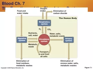
Cardiovascular System • Main function: Transportation • Blood = transport vehicle • Heart = pump • Blood vessels = network of tubes
Anatomy of Heart • Size of fist • Weight < 1 lb. • Apex points toward left hip • Flanked by lungs • Surrounded by pericardium (double-walled sac)
Layers of the Heart Wall • Epicardium – outer layer (pericardium) • Myocardium – cardiac muscle • Endocardium – endothelium lines chambers Heart chamber
Heart Chambers • Atrium (R & L): receive blood (entryway) • Ventricle (R & L): pump blood out • Septum: wall between atria & ventricles • Valves: prevent backflow of blood Right Side Left Side
Double Circulation Loop • Pulmonary circuit: blood to/from lungs • Systemic circuit: blood to/from all body tissues
Pathway of Blood Through Heart • To Left Atrium: • 4 Pulmonary veins (lungs to heart) • To Right Atrium: • Superior vena cava (above diaphragm) • Inferior vena cava (below diaphragm) • Coronary sinus (from myocardium) Mitral (bicuspid) valve Tricuspid valve • Left Ventricle: • Aorta(to body) • Coronary arteries • Right Ventricle: • 2 Pulmonary arteries(to lungs) Aortic valve Pulmonary valve
Right Ventricle Left Ventricle • Pulmonary circuit = low pressure • Systemic circuit = high resistance to blood flow • More powerful pump • 3X as thick as right ventricle
Coronary Circulation Coronary arteries Coronary veins
Heart Valves • Atrioventricular (AV) valves (tricuspid, bicuspid) • Semilunar valves (pulmonary, aortic)
Heart Valves • Chordae tendineae: anchors valve flaps in their closed position
Anatomy of the Heart Web Activity http://www.wisc-online.com/Objects/ViewObject.aspx?ID=ap12504
Warm-Up • Draw the human heart and the main blood vessels in/out of the heart. • Label the following on your diagram: • 4 chambers • 4 valves • All blood vessels going into/out of heart • Using a blue pencil, indicate oxygen-poor blood flow • Using a red pencil, indicate oxygen-rich blood flow
Heart Rhythm • Cardiac muscle cells can contract spontaneously and independently • Regulation of heart activity: • Autonomic nervous system • Epinephrine, thyroxine: heart rate • Low Ca2+ levels: heart rate • Intrinsic conduction system • Built into heart tissue & sets basic rhythm • Pacemaker = Sinoatrial (SA) Node
Intrinsic conduction system Sequence of action: • Sinoatrial (SA) node – right atrium • Generates impulses Starts each heartbeat • Atrioventricular (AV) node – between atria & ventricles • Atria contract • Bundle of His (or AV bundle) • Bundle branches – interventricular septum • Purkinje fibers – spread within ventricle walls • Ventricles contract
Electrocardiogram (ECG/EKG) • Records the electrical activity of the heart • Electrocardiograph: graphic record of heart activity
How to read an ECG • P wave: atria contact • QRS complex: ventricles contract • T wave: ventricles relax