Download
1 / 28
290 likes | 329 Vues
Explore the molecular factors in amyloid formation, protein misfolding, and neurodegenerative diseases such as Alzheimer's. Learn about the Prion replication cycle and mechanisms of prion progression.

E N D
Biochemistry of neurodegenerative diseasesand prions Alice Skoumalová
α-helix β-sheet Conformational change • the starting point is the natural protein folded in the native and active conformation • normal protein is rich in α-helix conformations (folded structure) • the end-point is the same protein adopting prevalent β-sheets structure • it is disease-associated protein (misfolded structure) Aggregation Gain of toxic activity Neurodegenerative diseases Loss of biological function
DNA Ubiquitin Ribosome RNA ATP Chaperones Native protein Misfolded protein Aggregate/fibrillar amyloid Chaperones Proteasome Accumulation (Amyloidoses) Degraded protein Gain of toxicity (Alzheimer disease) Loss of protein function (Cystic fibrosis)
Amyloid fibril structure Straight, unbranched, diameters in the range of 8-16nm Composed of two to six protofilaments Rich in a type of β-sheet structure (the β-sheets are perpendicular to the fibril axis and bind together by the hydrogen bonds)
Molecular factors in amyloid formation Protein misfolding is central to amyloid formation Protein stability- the resistance of the folded conformation to misfolding- is an important factor in determining susceptibility to amyloid formation Destabilizing factors: 1. Extreme environments in the body, such as acidic cell compartments 2. Proteolytic removal of a portion of a protein by an endogenous protease 3. Mutations that alter the primary structure (many of the amyloid diseases involve amino acid substitutions in an amyloid precursor protein) 4. Interactions with lipid bilayers (Aβ)
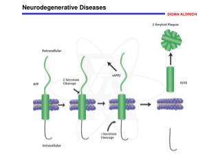
Histopathological hallmarks of Alzheimer disease • Neurofibrillary tangles • hyperphosphorylated protein tau • Amyloid (senile) plaques • protein β-amyloid (Aβ) fibrils • the abnormal processing of APP (β-amyloid precursor protein)
Amyloid cascade hypothesis of Alzheimer disease Missense mutations in APP, PS1, or PS2 genes Increased Aβ42 production and accumulation Aβ42 oligomerization and deposition as diffuse plaques Subtle effects of Aβ oligomers on synapses Microglial and astrocytic activation (complement factors, cytokines) Progressive synaptic and neuritic injury Altered neuronal ionic homeostasis; oxidative injury Altered kinase/phosphatase activities tangles Widespread neuronal dysfunction and cell death with transmitter deficits Dementia
Prions Prions are proteinaceous transmissible pathogens responsible for a series of fatal neurodegenerative diseases (in humans, Creutzfeld-Jakob disease and kuru, in animals, bovine spongioform encephalopathy) A prion (proteinaceous infectious particle, analogy for virion) is a type of infectious agent that does not carry the genetic information in nucleid acid! Prions are proteins with the pathological conformation that are believed to infect and propagate the conformational changes of the native proteins into the abnormally srtructured form
PrPC The normal protein is called PrPC (for cellular) is a transmembrane glycoprotein (neurons, lymphocytes); its function is unknown; it binds Cu2+ (regulation its homeostasis) has dominant secundary structure α-helix is easily soluble is monomeric and easily digested by proteases is encoded by a gene designated PRNP located on the chromosome 20 PrPSc The abnormal, disease-producing protein is called PrPSc (for scrapie) has the same amino acid sequence(primary structure) has dominant secundary structure β-sheets is insoluble is multimeric and resistant to digestion by proteases When PrPSc comes in contact with PrPC, it converts the PrPC into more of itself These molecules bind to each other forming aggregates
Molecular models of the structure of: PrPC PrPSc Predominantly α-helix (3) β-sheets (40%), α-helix (30%)
Replication cycle The presence of an initial PrPSc: exogenous (infectious forms) or endogenous (inherited or sporadic forms) This first prion will initiate PrPSc accumulation by sequentially converting PrPC molecules into PrPSc in replication cycle PrPSc molecules aggregate
Prion diseases: rare neurodegenerative disorders (one person per million) 1. Sporadic (85 %) In the sixth or seventh decade, rapidly progressive (death in less than a year) Creutzfeldt-Jakob disease (CJD) 2. Familial (inherited-15%) Mutations in the PrP gene that favour the transition from the cellular form to the pathological form of PrP Gerstmann-Straussler-Scheinker disease (GSS), fatal familial insomnia (FFI) 3. Transmissible (rare; a source of great concern) In the laboratory, disease can be readily transmitted to mice by intracranial injection of brain homogenate taken from prion-infected animals Propagation of kuru disease in New Guinea natives (ritualistic cannibalism) Recently, it has been discovered that BSE had been transmitted to humans in Europe after consumption of infected beef, producing a variant of the CJD called vCJD
Transmissible spongioform encephalopathy (TSE)=prion disease is a group of progressive conditions that affect the brain and nervous system of humans and animals and are transmitted by prions The pathology: vacuolar degeneration, neuronal loss, astrocytosis and amyloid plaque formation The clinical signs: loss of motoric functions (lack of coordination, ataxia, involuntary jerking movements), personality changes, depression, insomnia, confusion, memory problems, dementia, progressive tonic paralysis, death Definitive diagnostic test: biopsy of brain tissue There is no cure
Prion transmission 1. Direct contact with infected tissues CJD has been transmitted: -to patients taking injections of growth hormone harvested from human pituitary glands -from instruments used for brain surgery (prions can survive the autoclave sterilization process) -in corneal grafts -in electrode implants 2. Consumption of affected tissues Kuru was transmitted through cannibalism in Papua New Guinea Humans can contract the disease by consuming material from animals infected with the BSE (vCJD) How can prions make their way through the gut and into the brain? Proteins normally are digested down to amino acids in the gut Hypothesis: They circumvent the normal process of intestinal absorption by passing into the the Gut-Associated Lymphoid Tissue (GALT)
Prion strains =subclassification of prions following the structural characteristics that define their pathological profile The various prion diseases differ in incubation times and neuropathologic profiles, although in all cases the same misfolded protein (PrPSc) is responsible The existence of various prion strains: although they are chemically identical, they differ in their exact conformations The structural characteristics of individual prion strains are propagated via the replication cycle These characteristics appear to direct the precise tissue targeting, incubation time, and pathogenesis of the strain Consumption of BSE-infected meat BSE prion Variant CJD: younger cohort, distinct clinical characteristics vCJD prion is identical to BSE prion Classical CJD: older cohort CJD prion is different
Strains can be differentiated by characteristic incubation periods and neuropathology • distinct biochemical characteristics Properties of a single strain may be retained after passage in a range of different species -when re-isolated in the original host
Conformational selection and transmission barriers A wide range of PrPSc conformations A subset is compatible with each individual PrP Transmission between species with same conformations
Therapeutic strategies 1. Compounds can be designed to specifically disrupt the replication cycle of the PrPSc Design of such compounds had proven successful in cell-based models but must now be extended to animal models and human clinical trials 2. Vaccine design: The abnormally folded proteins expose a side chain of amino acids which the properly folded protein does not expose. Antibodies specifically coded to this side chain amino acid sequence stimulate an immune response to the abnormal prions 3. Design of peptides that break the β-sheet structures 4. Gene therapy: modification of the prion gene Genetic engineering research: cattle lacking a necessary gene for prion production - thus theoretically making them immune to BSE (December 2006)
Summary Conformational defect of proteins - protein misfolding diseases - neurodegenerative diseases Oligomeric aggregates - neurotoxic The mechanism: oxidative stress, disruption ion homeostasis The prions are proteins that carry information for self-reproduction (contradict the central dogma of modern biology) The prions are expressed in cells of healthy humans and animals; their abnormal conformations (PrPSc) are insoluble, resistent to digestion and aggregate The PrPSc attacks the native prion PrPC, changes its conformation into an abnormal form and causes an exponential production of insoluble proteins; they aggregate and form the fibrillar structure Prion disease are rare fatal degenerative disorders; a portion of them can be transmitted; this mechanism is not clear (e.g. transmision of BSE to human)