Functional and structural imaging in neurodegenerative diseases
380 likes | 634 Vues
Functional and structural imaging in neurodegenerative diseases. Caroline Sage Promotor: Prof. Dr. Stefan Sunaert Co-promotor: Prof. Dr. Wim Robberecht. Overview. Introduction Aims and methods Results Future directions. Overview. Introduction Aims and methods Results Future directions.

Functional and structural imaging in neurodegenerative diseases
E N D
Presentation Transcript
Functional and structural imaging in neurodegenerative diseases Caroline Sage Promotor: Prof. Dr. Stefan Sunaert Co-promotor: Prof. Dr. Wim Robberecht
Overview • Introduction • Aims and methods • Results • Future directions
Overview • Introduction • Aims and methods • Results • Future directions
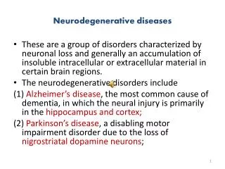
Introduction • Neurodegenerative diseases • Alzheimer’s disease • Parkinson’s disease • Multiple sclerosis • Huntington’s disease • Pick’s disease • Prion diseases • Amyotrophic lateral sclerosis
Introduction • Amyotrophic Lateral Sclerosis (ALS) • Cause is poorly understood • 5-10% familial ALS (fALS) • 90-95% sporadic ALS (sALS) • Loss of motor neurons (MN) • Upper MN signs • Lower MN signs • Spectrum disease?
Introduction • Research in ALS • Cell cultures & molecular research • Neuronal cells: motor neurons • Non-neuronal cells: astrocytes, oligodendrocytes, microglia • Agents for survival and neuronal protection (VEGF,...) • Animal studies • Mutant SOD1 mice and rats: overexpression of mutant SOD1 • Pathological mechanisms: glutamate excitotoxicity, impaired axonal transport,... • Rescue experiments • Human studies • Ex-vivo: autopsy of brain and/or spinal cord • Tissue studies: blood analysis, CSF analysis • In-vivo: PET, TMS, 1H-MRS, MRI Pubmed search dd 26/06/2007: 4568 scientific publications, in English, over the last 10 years!
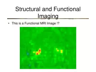
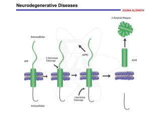
Introduction • Magnetic resonance imaging (MRI) in ALS • Conventional MRI • PD/T2w/FLAIR: non specific markers (Cheung et al., 1995; Hecht et al., 2001; Hecht et al., 2002) • T1w: loss of GM volume and to a lesser degree also loss of WM volume, especially in patients with cognitive deficits (Ellis et al., 1999; Abrahams et al., 2005; Grosskreutz et al., 2006) • Functional MRI (fMRI) • Motor tasks: recruitment of motor and non-motor areas in ALS patients (Konrad et al., 2002; Schoenfeld et al., 2005) • Cognitive tasks: cognitive deficits in ALS patients, especially in ALS patients with concomittant frontotemporal lobe dementia (Abrahams et al., 2006) • Diffusion tensor imaging (DTI) • Impairment of the corticospinal tract: reduction of FA and/or increase of Dav (Ellis et al., 2001; Toosy et al., 2003; Graham et al., 2004; Hong et al., 2004; Sach et al., 2004; Abe et al., 2005)
Overview • Introduction • Aims and methods • Results • Future directions
Aims • Research questions • Are there structural MRI changes in the brain of ALS patients? • Are there functional MRI changes in the brain of ALS patients? • Search for radiological correlates of structural and/or functional deficits in ALS patients by comparing ALS patients with a group of healthy age- and sex-matched controls • Design scan protocol of different tests for use in clinical settings • Improve diagnosis • Provide prognosis • Monitor newly developed therapies
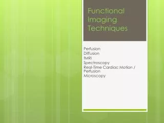
Neuronal function Cerebral vasoreactivity Cerebral vasculature/perfusion WM architecture fMRI vasoreactivity (VASC) fMRI motor tasks Dynamic contrast-enhanced T2*w imaging (PWI) Diffusion tensor imaging (DTI)
Overview • Introduction • Aims and methods • Results • Future directions
DTI - introduction • DTI • Diffusion Tensor Imaging • Assess Brownian motion of water molecules isotropy free diffusion restricted diffusion anisotropy
DTI - introduction • Data acquisition • Apply magnetic field gradients in multiple non-collinear directions during MRI data acquisition -> ° signal loss due to diffusion (Stejskal and Tanner, 1965) • Determine diffusion coefficient D in each voxel by varying b-value • In case of highly ordered structures: model diffusion by estimation of diffusion tensor D using multivariate fitting S = S0 e-bD
Dxx Dxy Dxz Dyx Dyy Dyz Dzx Dzy Dzz λ1 0 0 0 λ2 0 0 0 λ3 l1 l2 l3 DTI - introduction non diffusion-weighted image (b0) + ≥ 6 diffusion weighted images
3((l1-l2)2+ (l2-l3)2+ (l1-l3)2) 2(l12+ l22+ l32 ) l1 + l2 + l3 3 DTI - introduction • Derive quantitative diffusion parameters • Dav : amount of directionally averaged diffusion (in mm²/s) • FA : scalar measure of amount of anisotropy (0 = isotropic; 1 = diffusion in 1 specific direction only) Dav = FA =
DTI - introduction Mori et al., 1999
DTI - aim • Study white matter integrity in the brain of ALS patients by means of DT-MRI • Fibertracking of CST • Spatial interpolation of tract data • Voxel-based analysis of whole brain white matter • Correlation of disease severity with diffusion parameters • Quantitative comparison of diffusion parameters between ALS patients and controls • FA • Dav
DTI - material & methods • Subjects • Patients (PA, n = 28) • Sex: 14 female, 14 male • Age = 58.9 +/- 11.8 years • ALS-FRS= 39.7 +/- 6.3 • Controls (CT, n = 26) • Age = 53.7 +/- 11.8years • Sex: 15 female, 11 male • Imaging (3T) • DTI • 16 directions; b= 800 mm²/s; 2mm isotropic resolution • 3D-TFE
DTI - fibertracking • Check integrity of corticospinal tract (CST) • Motor part -> precentral gyrus • Sensory part -> postcentral gyrus • Reconstruct ‘mean’ CST + separate parts • Compare mean FA/Dav values between patients and controls
* * * * * * Precentral * * Postcentral n.s. * n.s. n.s. DTI - Fibertracking
DTI – interpolation of tract data • Assess local variation of FA/Dav values over course of CST • Interpolation of tract data to spatially ‘normalize’ tract data • Compare mean FA/Dav values between patients and controls
z DTI - Interpolation of tract data Interpolation of individual data in z-direction Measure FA/Dav over z-direction of interpolated data Select part of CST between pons and subcortical WM Tract data $ $ 76 « new » z-coordinates
* * FA * * Dav
DTI – voxel-based analysis • Assess WM integrity of whole brain • Normalize FA/Dav maps • Smooth warped maps • Voxel-by-voxel comparison of FA/Dav values in whole brain
X 28 ALS patients X 28 ALS patients X 26 controls X 26 controls t-test t-test T-value of test FA in PA < CT T-value of test Dav in PA > CT DTI – voxel-based analysis Test in each voxel
CST Orbitofrontal Prefrontal Hippocampal formations Insular regions Parietal regions WM underneath PMC WM underneath SMA p<0.05, FWE corrected DTI – voxel-based analysis
DTI - correlation analysis • Study effect of patients’ scores on ALS-FRS on FA/Dav • ALS-FRS: questionnaire of 12 questions to assess « functional integrity » of patients • Questions relate to day-to-day activities • max. score = 48 • Add individual score as a covariate in a voxel-based correlation analysis
A DTI - correlation analysis CST_ALSFRS_FA_positive Frontal_ALSFRS_FA_positive
DTI - summary • Significant impairment of CST in ALS patients • Limited to the precentral part of the CST • Mostly in cranial parts of the CST • White matter impairment is not limited to the motor system • Areas involved in voluntary motor control • Proprioceptive areas • Frontal/temporal/hippocampal structures • Strong correlation of ALS-FRS and FA • In CST • Especially in orbitofrontal cortex This study provides support for the view of ALS as being a multisystem degenerative disease, in which abnormalities of extra-motor play an important role in the in vivo physiopathology Sage et al., 2007
Overview • Introduction • Aims and methods • Results • Future directions
Future directions - DTI • Non-rigid coregistration of DTI data in cooperation with UZ Antwerpen (W. Van Hecke) • To reference • To atlas • Tract-based spatial statistics (TBSS, S. Smith et al., 2006)
Thank you for your attention Questions?