Structural and Functional Imaging
Structural and Functional Imaging. This is a Functional MRI Image !?. Structural and Functional Imaging. This is a structural MRI image (an “anatomical” image). Structural and Functional Imaging. What you really want is both images co-registered. Structural and Functional Imaging.

Structural and Functional Imaging
E N D
Presentation Transcript
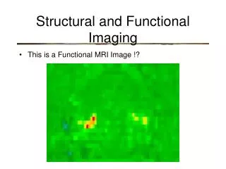
Structural and Functional Imaging • This is a Functional MRI Image !?
Structural and Functional Imaging • This is a structural MRI image (an “anatomical” image)
Structural and Functional Imaging • What you really want is both images co-registered
Structural and Functional Imaging • What you really want is both images co-registered • Why? What’s wrong with the functional image alone?
Structural and Functional Imaging • Functional images tend to be lower resolution and fail to convey spatial information Pixels
Structural and Functional Imaging • Structural images have finer (smaller) pixels Pixels
Structural and Functional Imaging • Brain scan images (CAT, PET, MRI, fMRI) are all made up of pixels (stands for picture elements) Pixels
Structural and Functional Imaging • “Slices” are assembled into “volumes” Pixels
Structural and Functional Imaging • Volumes are composed of “volume elements” or voxels Voxels
Structural and Functional Imaging • Another thing you want: the ability to tell other people where something is • “the activity was centered on voxel #653” will not work in a scientific journal
Structural and Functional Imaging • MRI anatomical spaces • Talairach Space: • Based on detailed analysis of one elderly woman • Talairach & Tournoux (1988) • Montreal Neurological Institute Template (MNI) • based on average of 152 different brains, each normalized to Talairach space • advantage: gyri and sulci are more representative • disadvantage: it’s blurry • MNI “Representative Brain” • the one brain from the 152 in the MNI Template set that is most like the average • advantage: it’s not blurry • disadvantage: it’s still just one person’s brain
Structural and Functional Imaging • Reasons for normalizing to standard stereotaxic space (templates) • two levels: within-subject and between-subjects
Structural and Functional Imaging • Within-Subject Reasons: • structural and functional volumes may not be coregistered due to • movement • distortion • results can be described in standard coordinates • data across sessions can be averaged
Structural and Functional Imaging • Between-Subject Reasons: • Volumes will not match because of variability across individuals • results can be described in standard coordinates • data across participants can be averaged
Preprocessing of Structural and Functional Images • Normalizing images to fit a standard template (e.g. Talairach) • Define Coordinate System using easily recognizable landmarks • Origin in the Anterior Commissure • y-axis connects AC and PC • x-axis perpendicular interhemispheric plane and through AC • z-axis perpendicular to x and y
The Talairach Coordinate System -y AC - PC line defines y-axis +y
The Talairach Coordinate System -y +x x-axis perpendicular to interhemispheric plane -x +y
The Talairach Coordinate System +z -y +x z-axis perpendicular to x-y plane -x -z +y
Structural and Functional Imaging • Cortical Flattening • Software such as BrainVoyager can “inflate” the cortex like a balloon so that sulci and gyri are “flattened” • functional data can be transformed with the same complex function • functional and structural data can be overlaid so that distribution on cortical sheet can be visualized