Download
1 / 55
670 likes | 1.48k Vues
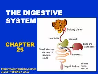
Figure 24.1 The Components of the Digestive System. Figure 24.1. Functions of the digestive system. Ingestion Mechanical processing Digestion Secretion Absorption Excretion. Figure 24.3 The Structure of the Digestive Tract. Figure 24.3. Movement of digestive materials.

E N D
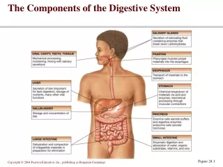
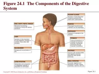
Figure 24.1 The Components of the Digestive System Figure 24.1
Functions of the digestive system • Ingestion • Mechanical processing • Digestion • Secretion • Absorption • Excretion
Figure 24.3 The Structure of the Digestive Tract Figure 24.3
Movement of digestive materials • Visceral smooth muscle shows rhythmic cycles of activity • Pacemaker cells • Peristalsis • Waves that move a bolus • Segmentation • Churn and fragment a bolus
Figure 24.4 Peristalsis Figure 24.4
Control of the digestive system • Movement of materials along the digestive tract is controlled by: • Neural mechanisms • Parasympathetic and local reflexes • Hormonal mechanisms • Enhance or inhibit smooth muscle contraction • Local mechanisms • Coordinate response to changes in pH or chemical stimuli
Figure 24.5 The Regulation of Digestive Activities Figure 24.5
The mouth opens into the oral or buccal cavity • Its functions include: • Analysis of material before swallowing • Mechanical processing by the teeth, tongue, and palatal surfaces • Lubrication • Limited digestion
The tongue • primary functions include: • Mechanical processing • Assistance in chewing and swallowing • Sensory analysis by touch, temperature, and taste receptors
The pharynx • Common passageway for food, liquids, and air • Lined with stratified squamous epithelium • Pharyngeal muscles assist in swallowing • Pharyngeal constrictor muscles • Palatal muscles
Histology of the esophagus • Distinctive features of the esophageal wall include • Nonkeratinized, stratified squamous epithelium • Folded mucosa and submucosa • Mucous secretions by esophageal glands • A muscularis with both smooth and skeletal muscle portions • Lacks serosa • Anchored by an adventitia
Figure 24.10 The Esophagus Figure 24.10a-c
Figure 24.11 The Swallowing Process Figure 24.11a-h
Functions of the stomach • Bulk storage of undigested food • Mechanical breakdown of food • Disruption of chemical bonds via acids and enzymes • Production of intrinsic factor
Digestion and absorption in the stomach • Preliminary digestion of proteins • Pepsin • Permits digestion of carbohydrates • Very little absorption of nutrients • Some drugs, however, are absorbed • Mucous secretion containing several hormones
Figure 24.12 The Stomach Figure 24.12b
Figure 24.13 The Stomach Lining Figure 24.13a, b
Figure 24.13 The Stomach Lining Figure 24.13c, d
Histology of the stomach • Gastric glands • Parietal cells • Intrinsic factor, and HCl • Chief cells • Pepsinogen • Pyloric glands
Figure 24.14 The Secretions of Hydrochloric Acid Figure 24.14
Figure 24.15 The Phases of Gastric Secretion Figure 24.15a
Figure 24.15 The Phases of Gastric Secretion Figure 24.15b
Figure 24.15 The Phases of Gastric Secretion Figure 24.15c
Small intestine • Important digestive and absorptive functions • Secretions and buffers provided by pancreas, liver, gall bladder • Three subdivisions: • Duodenum • Jejunum • Ileum • Ileocecal sphincter • Transition between small and large intestine
Figure 24.16 Regions of the Small Intestine Figure 24.16a
Histology of the small intestine • Plicae • Transverse folds of the intestinal lining • Villi • Fingerlike projections of the mucosa • Lacteals (involved in protein absorpbtion • Terminal lymphatic in villus • Intestinal glands • Lined by enteroendocrine, goblet and stem cells
Figure 24.17 The Intestinal Wall Figure 24.17a
Figure 24.17 The Intestinal Wall Figure 24.17b, c
Figure 24.17 The Intestinal Wall Figure 24.17d, e
Intestinal juices • Moisten chyme • Help buffer acids • Maintain digestive material in solution
Small Intestine • Duodenal glands (Brunner’s glands) • produce mucus, buffers • Ileum • aggregated lymphoid nodules (Peyer’s patches)
Intestinal movements • Peristalsis • Segmentation • Gastroenteric reflexes • Initiated by stretch receptors in stomach • Gastroileal reflex • Triggers relaxation of ileocecal valve
The pancreas • Pancreatic duct penetrates duodenal wall • Endocrine functions • Insulin and glucagons • Exocrine functions • Majority of pancreatic secretions • Pancreatic juice secreted into small intestine • Carbohydrases • Lipases • Nucleases • Proteolytic enzymes
Figure 24.18 The Pancreas Figure 24.18a-c
The liver • Performs metabolic and hematological regulation and produces bile • Histological organization • Lobules containing single-cell thick plates of hepatocytes • Lobules unite to form common hepatic duct • Duct meets cystic duct to form common bile duct
Figure 24.19 The Anatomy of the Liver Figure 24.19a
Figure 24.19 The Anatomy of the Liver Figure 24.19b, c
Figure 24.20 Liver Histology Figure 24.20a, b
The gallbladder • Hollow, pear-shaped organ • Stores, modifies and concentrates bile PLAY Animation: Accessory Organ
Figure 24.21 The Gallbladder Figure 24.21a, b
Coordination secretion and absorption • Neural and hormonal mechanisms coordinate glands • GI activity stimulated by parasympathetic innervation • Inhibited by sympathetic innervation • Enterogastric, gastroenteric and gastroileal reflexes coordinate stomach and intestines
Figure 24.22 The Activities of Major Digestive Tract Hormones Figure 24.22
Functions of the large intestine • Reabsorb water and compact material into feces • Absorb vitamins produced by bacteria • Store fecal matter prior to defecation
Figure 24.23 The Large Intestine Figure 24.23a
Figure 24.23 The Large Intestine Figure 24.23b, c
The rectum • Last portion of the digestive tract • Terminates at the anal canal • Internal and external anal sphincters
Histology of the large intestine • Absence of villi • Presence of goblet cells • Deep intestinal glands
Physiology of the large intestine • Reabsorption in the large intestine includes: • Water • Vitamins – K, biotin, and B5 • Organic wastes – urobilinogens and sterobilinogens • Bile salts • Toxins • Mass movements of material through colon and rectum • Defecation reflex triggered by distention of rectal walls
Figure 24.25 The Defecation Reflex Figure 24.25
Processing and absorption of nutrients • Disassembles organic food into smaller fragments • Hydrolyzes carbohydrates, proteins, lipids and nucleic acids for absorption