Anaesthesia for MRI
Anaesthesia for MRI . Dr. S. Parthasarathy MD., DA., DNB, MD ( Acu ), Dip. Diab.DCA , Dip. Software statistics PhD ( physio ). Definition .

Anaesthesia for MRI
E N D
Presentation Transcript
Anaesthesia for MRI Dr. S. Parthasarathy MD., DA., DNB, MD (Acu), Dip. Diab.DCA, Dip. Software statistics PhD (physio)
Definition • MRI is a noninvasive diagnostic technique that uses magnetic properties of atomic nuclei to produce high-resolution, multi planar cross- - sectional images of the body. • Hydrogen is the atom most often used for imaging.
Technology generates a high-density static magnetic field, which is always present. Field strength is measured in Tesla (T) most scanners use 0.5-1.5 T magnets (about 10,000 times the Earth's magnetic field).
Physics • some atoms within human body possess an unpaired proton, eg hydrogen atom • alignment of these nuclei are random • subjected to a strong electromagnetic field, they align themselves with the field • The rate of the alignment depends on the type of nucleus/ element • emitted signal depends on the molecular properties of the tissue
Various equipments and diffwerent place MRI – lumbar, knee, neck
The magnetic field decreases as distance from the scanner increases—beyond the 0.5 mT boundary or outside the scan room can be considered safe. This boundary is known as the 5-Gauss Line. 1,000 Gauss equals 1 T.
MRI is a valuable non invasive imaging technique. • MRI produces a high quality images of the body in cross-section and in three dimension • MRI is particularly useful for the imaging of soft tissues eg CNS • Revolutionized ortho, neuropractice
Hiccups with MRI • Noisy and unfamiliar environment • Motion interferes with images quality • MRI scan takes up to an hour • Hence patients may need anaesthesia
few indications for anesthesia • Infants and children • Patients with learning difficulties • Patients with seizure disorders • Patients with claustrophobia • Critically ill-patients • SL CCC – pneumonic
strong magnetic field- hazards • the attraction of ferromagnetic objects to the magnetic field of an MRI. • risk of dislodgement of implanted metallic objects (i.e., pacemakers, vascular clips, automatic implantable cardioverter defibrillators, mechanical heart valves, and implanted infusion pumps), • Orthodontic braces and dentures and tattoos can degrade the image quality significantly.
Other problems • Injury to patients, personnel, and equipment -- from propelled ferromagnetic objects brought into the magnetic field. • What are those objects ?? • gas cylinders, pens, keys, laryngoscopes, scissors, stethoscopes, paper clips, vials, and needles • malfunction of electronic equipment (such as monitors and infusion pumps)
MRI - four zones: • Zone I is the public zone • Zone II is the reception • Zone III – control room • Zone IV – scanner room
Personnel safety • no significant deleterious effects to patients or health care professionals from exposure to the static magnetic field of an MRI. • Pregnant patients have undergone MRI safely during all stages of pregnancy. Nevertheless, caution is advised. • Think USG only • the simple rule that ‘nothing enters the scan room except the patient.’
MR safe’ or ‘MR compatible’. • it presents no safety hazard to patients or personnel. guarantee that it will function normally and not interfere with the correct operation of the MR imaging equipment, with degradation of image quality. • MR compatible is MR safe, functions normally in the MR environment
Monitors • MR compatible pulse oximeters must use fibreoptic cabling to avoid burns • Special ECG electrodes and cables are required. • Padding should be placed between cables and the patient’s skin and the avoidance of loops in cables within the scanner
Some hiccups • Aortic blood flow in a magnetic field generates currents that result in significant artefact in the ST-T region of the ECG complex. • Delay of up to 20 seconds in obtaining the capnograph signal due to the length of the • sampling tubing. • The need for acoustic protection during MR imaging will necessitate the use of ear-plugs or ear defenders.
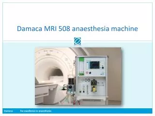
Separate machine • A MR compatible anaesthetic machine should be located within the scanning room. • Only MR compatible vaporisers and gas cylinders must be used on anaesthetic machines within the scanner room.
Anaesthesia team • Consultant • Team • Staff • Radio staff • Protocol • Beware -- credit cards, cassette tapes, or floppy disks ??
MRI room is for radiologists – not for anaesthesiologists • MRI suites have been designed without the • consideration for anesthetic needs, such as pipeline gases, suction, • bulkiness of the unit, • the patient is often far from the anesthesiologist, • access to the airway is limited. • Intravenous lines, anesthesia circuits, oxygen tubings, monitoring cables must be of sufficient length to reach the patient deep within the scanner.
Acoustic problems • Noise levels above the safe level of 85 decibels can be produced during MRI due to the rapid switching of the gradient. • Staff working in MRI units should protect themselves by remaining in the MR control room during sequence acquisition, or by wearing earplugs • All patients should be given ear protection, regardless of if they are awake or anaesthetised.
Practical considerations • Induction area adjacent to but outside the scan room (beyond the 0.5 mT boundary - 5-Gauss Line) equipped with a compact conventional anesthesia machine and monitoring. • Piped gases, scavenging, and suction in both the induction area and the control room. • Nonmagnetic gurney for patient transfer into scanner. – stretcher routine ??
Practical considerations • Respiratory gas/agent side-stream analyzer with capnograph display fitted with an extended sampling tube (increases the response time by 5-10 s). • MRI-compatible pulse oximeter (fiber-optic patient probe, and shielded cable).
Practical considerations • Compact (e.g., wall mounted) anesthesia machine and ventilator in the control room with a 10-m co-axial (Bain) or circle breathing system. • ECG with MRI compatible (carbon fiber) patient leads and electrodes. • NIBP machine with an extended hose, nonmetallic connectors, and a range of cuffs.
It is painless !! • Remember • MRI is painless procedure • Preop investigations • Nothing special depends upon the disease • Plates, pacemaker etc
Children • oral midazolam (0.5mg/kg, maximum 10 mg) in a small amount of cherry or strawberry syrup. • Disabled children do not need higher doses of sedatives but are three times more at risk of hypoxia under sedation
Anaesthetic options • Options • Sedation ,TIVA, Controlled GA
Alternative to fasting • A very safe and simple technique for newborns is the ‘feed and scan’ technique in which children are fed and one has to wait until the young patient falls asleep. • unpredictable ‘induction times’ and the high failure rates of the scanning procedure
Sedation – options • Other sedative options • Oral chloral hydrate • Dose - 25 and 100 mg/kg • nausea and vomiting, long recovery times and postoperative agitation • Oral melatonin
Other sedative techniques • Pentobarbital – oral or rectal – 3-6 mg/kg • Dosing is 1–1.5 mg/kg when applied intravenously or 4–5 mg/kg when injected intramuscularly. Onset time is 1–3 min, and duration is 15–30 min • What is this drug ?? • Ketamine
Propofol • Propofol seems to be a perfect drug for sedation because it is effective, has a short recovery time and can easily be titrated to the required sedation level. • Dosing is normally 2–5 mg/kg/h intravenous
Other sedatives • Dexmed • A loading dose of 2–3mic.g/kg over 10 min followed by 1–2mic. g/kg/h as an infusion for sedation maintenance is recommended. • Midazolamused alone is not suitable for MRI sedation as its duration is too short for a successful procedure of 20–30 min • Can be combined – be careful
When sedation, when GA ?? • > 3 years , 10 kg , no comorbidities • Consider sedation • < 3 years < 10 kg , lot of co illness – GA • Some may sleep without drugs - students !!
GA • Control room :: Prior to induction, patients should be placed on a non-ferrous trolley (without oxygen cylinders) so that they can be safely transferred to the MR room once they are anaesthetised. • Induction agents includes sodium thiopental (5mg/kg) , propofol (2-3mg/kg) • Secure airway with LMA • Second metal check ; remove stethoscope, needles , oxygen cylinder from the trolley • Transfer patient to MRI room
Procedure • Transfer patient onto MRI scanner • Check to ensure the airway is secured • Maintenance of anaesthesia may be either inhalational or intravenous • TIVA is an acceptable technique for MRI
TIVA • Propofol infusion ( 6-10mg/kg/hr) for patient • Infusion pump should be keep in control room, while connecting lines are passed through waveguide. • Ear protection before commencing scanning • Anaesthetists monitor the patient from control room
Recovery • Once study has been completed, patient should be remove from the scanner, and be woken up and recovered in a suitable recovery area • Usually there is no need for analgesics • Patients for MRI scan are usually day case
Contrast agents • The most commonly used intravenous MR contrast agent is gadolinium dimeglumine. • contrast-enhanced MR angiography. • doses of 0.2 ml/kg and has minor side effects including nausea, vomiting and pain • on injection. • Nephrotoxic ??
Maintenance of body temperature • More in infants • No active heating necessary. • Once the study has been completed, the patient should be removed from the scanner and be woken up and recovered in a suitable recovery area. • Analgesia is unlikely to be required for MRI scanning,
EMERGENCIES IN THE MRI SUITE • the presence of a strong magnetic field • risk of projectiles, • restricted access imposed by the MRI scanner. The patient should be removed from the magnetic field as quickly as possible and transferred to the induction room, which should be close to the scanner and will contain the necessary anaesthetic and resuscitation equipment and drugs
INTENSIVE CARE PATIENTS REQUIRING MRI • multiple drug infusions • extensions of adequate length or wave guides • higher standard of monitoring. • MR compatible monitors – must • Pulmonary artery catheters with conductive wires in contact with heart muscle and epicardial pacing wires – removal plannned
INTENSIVE CARE PATIENTS REQUIRING MRI • Many tracheostomy tubes are not MR compatible and will need to be changed prior to the examination. • pre-MR X-ray screen – history vague • The pilot balloons of cuffed tracheal tubes may contain a small ferromagnetic spring will need to be taped securely away from the area being scanned
Health staff • long-term repeated exposure to strong magnetic fields has a harmful effect on the human body. • current recommendations suggest that a time-weighted average of 200mT over any 8-hour period should not be exceeded by healthcare personnel. • Ideally all staff should vacate the MRI examination room whilst the scan is in progress.
Summary • MRI – introduction and physics • Which patients need anesthesia ?? • Preop • Anesthesia • Emergence • Special !!