Exploring the Human Brain: Structure, Evolution, and Dominance
840 likes | 891 Vues
Dive into the complexities of the human brain, its structure, evolution, and significance in determining intelligence and dominance in the animal kingdom. Learn about functional areas, cerebral hemispheres, and the corpus callosum's role in information transfer.

Exploring the Human Brain: Structure, Evolution, and Dominance
E N D
Presentation Transcript
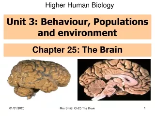
Higher Human Biology Unit 3: Behaviour, Populations and environment Chapter 25: The Brain Mrs Smith Ch25 The Brain
Learning Intentions Success Criteria To examine the workings of the brain and the nervous system. • Outline the structure of the human brain with reference to; • Size • The cerebrum and its convoluted surface • Localisation of function in discrete areas of the cerebrum • The relationship between size of a discrete are and the function carried out • The importance of the corpus callosum in transferring information between two hemispheres Mrs Smith Ch25 The Brain
The brain is a very complex organ not fully understood by scientists Variables Mrs Smith Ch25 The Brain
FYI: The Brain • Weighs 1300 - 1400g • Made up of about 100 billion neurons. • “The most complex living structure on the universe” Society for Neuroscience • Makes us who we are. Mrs Smith Ch25 The Brain
“No man's knowledge here can go beyond his experience”. • John Locke was an influential philosopher from the 17th century. He has provided many important ideas and bases on philosophy, one of which was his theory of personal identity. Locke believed all true knowledge came from the senses and human experience. • Basically each of us are a combinations of all the experiences we ever had and how we perceive these experiences. This can only happen with a brain to perceive, process, store, these experiences “We are like chameleons, we take our hue and the colour of our moral character, from those who are around us.” Mrs Smith Ch25 The Brain
The brain & size of the brain • The brain is a large organ composed of billions of nerve cells (neurones). • Compared with other animals, the human brain is disproportionally large, relative to body size. Mrs Smith Ch25 The Brain
Why is it appropriate to say the weight of the brain determines intelligence? AnimalWt. of BrainBrain/Body wt ratio Whale 15 lbs 1/10000 Elephant 3 lbs 1/1000 Human 3 lbs 1/50 • The human brain is more developed and has a larger weight in proportion to total body weight. Mrs Smith Ch25 The Brain
Evolution of the Brain: Fossil evidence Mrs Smith Ch25 The Brain
Increase in Brain Size Fossil evidence has shown that the human brain has increased in capacity (volume of skull occupied by the brain) over a fairly rapid evolutionary timescale. Mrs Smith Ch25 The Brain
Apes • When compared with the brains of modern apes, the human is found to be approximately three times larger. • Humans have much larger centres responsible for higher mental faculties such as intelligence, speech, hearing and sight. • An apes area controlling speech is so small and poorly developed it is impossible to teach an ape to speak. Mrs Smith Ch25 The Brain
Image source: http://www.nature.com Ape Brains Compared to the brain of modern apes the human brain is about 3 times larger. Humans have much larger centres responsible for higher mental faculties such as intelligence, speech, hearing and sight. An apes area controlling speech is so small and poorly developed it is impossible to teach an ape to speak. . Mrs Smith Ch25 The Brain
Dominant Species • Compared to other animals humans are physically weak. • However humans have become a dominant species on earth because the human brain has: • a larger size • complex internal development • complex organisation Mrs Smith Ch25 The Brain
Brain Structure:~ Revision Mrs Smith Ch25 The Brain
Cerebrum Structure • Controls conscientious thought, voluntary actions, determines personality etc Mrs Smith Ch25 The Brain
The Cerebrum: localisation of function The cerebral hemisphere has several distinct regions each with a particular function. Mrs Smith Ch25 The Brain
2 sides of the brain are joined by the corpus callosum - a large bundle of nerve fibres. This allows information to be transferred from one side to the other. Cerebrum: Con’t • Largest part of human brain • Split into 2 halves called cerebral hemispheres Mrs Smith Ch25 The Brain
Each side of the brain controls the other side of the body Mrs Smith Ch25 The Brain
Cerebrum: Grey and white matter • inner cerebrum • made of nerve cell fibres • surface of cerebrum • made of nerve cell bodies Mrs Smith Ch25 The Brain
The cerebrum’s surface is convoluted (folded) to give it a large surface area allowing many cell bodies to be close together. This maximises the potential for interconnections and the transmission of messages. Cerebrum Cerebrum: Convolution Mrs Smith Ch25 The Brain
Task: Torrance-TYK pg 206 Qu 1-4 Mrs Smith Ch25 The Brain
Discrete Functional Areas The cerebrum has 3 main types of functional area, which are all discrete (they have their own function): • Sensory • Association • Motor View the Scholar animation: http://courses.scholar.hw.ac.uk/vle/scholar/session.controller?action=viewContent&contentGUID=2ba96ae7-1eff-e695-8b5d-5f08e2cc8533 Mrs Smith Ch25 The Brain
Roles of the 3 discrete functional areas Association areas Analyse & Interpret sensory impulses e.g. Make decisions Motor areas Receive info from association areas & send motor impulses to the effectors e.g. muscles Sensory areas Receive info as sensory impulses from body’s receptors e.g. touch receptors in skin and thermoreceptors in hypothalamus Mrs Smith Ch25 The Brain
Premotor association area Somatosensory association area Auditory association area Visual association area Association areas Mrs Smith Ch25 The Brain
Speech Each region of the left cerebral hemisphere is duplicated on the right cerebral hemisphere except speech. Each person only has one speech area. In 90% of people this is in the left cerebral hemisphere. Mrs Smith Ch25 The Brain
Motor area Association area Interconnections in the Brain Tiny nerve fibres interconnect the different areas of the brain. Messages constantly pass between them. Sensory area This allows the human brain to cope with several sensory impulses at once (sophisticated perception) They then cause more exchange of impulses between cerebral areas allowing a sophisticated response e.g. channel flicking e.g. singing & dancing Mrs Smith Ch25 The Brain
The motor area is one of the largest regions of each cerebral hemisphere. Each motor area consists of motor neurons which sends out impulses to bring about voluntary movement of skeletal muscles However, the size of the part of the motor area is not in proportion to the actual size of the body part. The size of the motor area is in proportion to the number of nerve endings in the body part Motor Area Mrs Smith Ch25 The Brain
Discovery of the motor area In 1870, Hitzig and Fritsch electrically stimulated parts of a dog's motor cortex. Depending on what part of the cortex they stimulated, a different part of the body contracted. When they destroyed this same small area of the cortex, the corresponding part of the body became paralysed. They concluded that every part of the body has a particular region of the primary motor cortex that controls its movement. Mrs Smith Ch25 The Brain
Motor or sensory area allocated to a particular body part is found to be in relative proportion to its mobility/sensory. e.g. the more mobile a part the larger the motor area. Mrs Smith Ch25 The Brain
Who is Homunculus? • Imaginary human whose body parts have been drawn in proportion to • Mobility and fine motor control • Sensory perception • The more control needed of a task, the larger the area of the brain that is required Mrs Smith Ch25 The Brain
Motor homunculus—larger parts of the brain control larger parts of the body such as the hand and mouth, which require a lot of “motor” or motion “signals.” That is, if the human body were to be built in proportion to its motor significance because of the brain power needed to “motor” them, the hands and mouth would be proportionally bigger. The motor area of the left cerebral hemisphere Motor homunculus "This model shows what a man's body would look like if each part grew in proportion to the area of the cortex of the brain concerned with its movement." View the Scholar animation: http://courses.scholar.hw.ac.uk/vle/scholar/session.controller?action=viewContent&contentGUID=8af4bea2-600b-deeb-baec-d7517d578e46 Mrs Smith Ch25 The Brain
Sensory homunculus—Similar to motor homunculus but it tells the brain how much power is needed for sensory perception of different body parts. The sensory area of the left cerebral hemisphere Sensory homunculus "This model shows what a man's body would look like if each part grew in proportion to the area of the cortex of the brain concerned with its movement." Mrs Smith Ch25 The Brain
Cerebellum • Attached to underside of brain • Unconscious fine control of voluntary muscle movement and balance Mrs Smith Ch25 The Brain
Medulla oblongata Medulla Oblongata • Connects brain to spinal cord • Unconscious co-ordination of basic functions – breathing, heart rate, digestion, reflex actions Mrs Smith Ch25 The Brain
Studying the Brain • The evidence that there is localisation of brain functions (i.e. that different parts of the brain have different functions) include: • Electroencephalograms (EEG’s) B. Brain Scans • Cat Scan • fMRI Scan Mrs Smith Ch25 The Brain
Brain Scans Speech involves several specific regions of the brain. These show up in brain scans as areas of high metabolic activity. FYI: You DON’T need to know the names of these areas!! View the Scholar animation: http://courses.scholar.hw.ac.uk/vle/scholar/session.controller?action=viewContent&contentGUID=c622c31f-213c-2853-d6e0-e311a44d055f Mrs Smith Ch25 The Brain
Studying the Brain- EEG Electroencephalograms A record of the cerebrum’s electrical activity. Electrodes are placed on the different regions of the scalp. They detect impulses which are displayed on a monitor. Different brain wave patterns show different levels of mental activity. Mrs Smith Ch25 The Brain
EEG wave patterns The more densely packed the spikes, the higher the level of electrical activity in the brain. EEGs are not very precise because they reflect the activity of many brain cells. Children, Sleeping adults EEGs can show abnormal patterns that indicate a problem (e.g. dense spikes are shown during epileptic attacks), but the EEG doesn’t show the area of the brain responsible. Infants, Sleeping adults Epilepsy Mrs Smith Ch25 The Brain
Often referred to as CAT (Computer Assisted Tomography) scans. These give a clear image of the brain without any surgery used mainly to diagnose abnormalities Can indicate areas of high metabolic activity – so can be used to determine which part of the brain is responsible for certain actions and emotions. This brain scan shows a tumour in pale blue. Image source: www.sciencemuseum.org.uk Studying the Brain: Brain Scans CAT SCANS Mrs Smith Ch25 The Brain
Studying the Brain: Brain Scans fMRI SCANS • Brain scans provide pictures of very active parts of the brain • The parts of the brain which are active show up as brightly coloured areas • The following diagrams show four fMRI (functional magnetic resonance imaging) brain scans obtained during a visual memory task. Mrs Smith Ch25 The Brain
Scan 1 In scan 1, a subject is asked to remember a face. Areas at the rear of the brain that process visual information are active during this task, as is an area in the frontal lobe. Mrs Smith Ch25 The Brain
Scan 2 In scan 2, the subject is asked to "think about this face." The hippocampus is activated. The hippocampus was already known to be important for memory, but these results show that this part of the brain is specifically active during the time when we are remembering new information. Mrs Smith Ch25 The Brain
Scans 3 and 4 • In scans 3 and 4, the subject was asked to compare another face to the remembered face. Some of the same visual areas are activated as during the initial memory task, but other areas, such as part of the frontal lobe, are involved in making a decision about the memory. Mrs Smith Ch25 The Brain
Split-brain studies • Split brain happens when a person’s corpus callosum has been cut. • Because of this exchange of information between cerebral hemispheres doesn’t occur. • Learn more............. • http://www.nobelprize.org/educational/medicine/split-brain/background.html • Play the split brain game................. • http://www.nobelprize.org/educational/medicine/split-brain/index.html Mrs Smith Ch25 The Brain
Split Brain Studies Visual Pathways – Normal situation When the corpus callosum is intact both hemispheres perceive all information from both eyes as each hemisphere quickly communicates so the whole picture is in view Each cerebral hemisphere only receives half the information of the visual field. Everything to the left Is represented by the right cerebral hemisphere Everything to the right is represented by the left cerebral hemisphere. Mrs Smith Ch25 The Brain