RRT Review
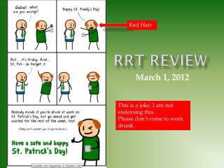
Gabe!. Red Hair. RRT Review. March 1, 2012. This is a joke. I am not endorsing this. Please don ’ t come to work drunk. MKSAP.

RRT Review
E N D
Presentation Transcript
Gabe! Red Hair RRT Review March 1, 2012 This is a joke. I am not endorsing this. Please don’t come to work drunk.
MKSAP • A 30-year-old woman develops bilateral pulmonary infiltrates and hypoxemia 48 hours after undergoing repair of multiple long-bone fractures. Her initial arterial blood gases are pH 7.48, PCO2 30 mm Hg, and PO2 45 mm Hg on 100% oxygen by nonrebreather mask. The patient is intubated and placed on mechanical ventilation. • The temperature is 38.3 °C (101.0 °F), the pulse rate is 100/min, the respiration rate is 28/min, and the blood pressure is 120/60 mm Hg. The patient weighs 60 kg (132.3 lb). Oxygen saturation is 83%. She is sedated, hemodynamically normal, and not using accessory muscles to breathe. There are bilateral inspiratory crackles. The ventilator settings are as follows: volume control mode, respiration rate 26/min, tidal volume 360 mL, positive end-expiratory pressure (PEEP) 5 cm H2O, FiO2 0.8. Arterial blood gases are pH 7.45, PCO2 33 mm Hg, and PO2 50 mm Hg.
MKSAP • Which of the following is the most appropriate next step in the management of this patient? • Increase FiO2 to 1.0 • Increase PEEP to 10 cm H2O • Increase respiration rate to 32/min • Increase tidal volume to 700 mL • Start vecuronium infusion
Please make your selection... • Increase FiO2 to 1.0 • Increase PEEP to 10 cm H2O • Increase respiration rate to 32/min • Increase tidal volume to 700 mL • Start vecuronium infusion
The patient has severe hypoxemic respiratory failure from the ARDS. The primary cause of hypoxemia is shunt, which will not correct with breathing 100% oxygen. Increasing positive end-expiratory pressure (PEEP) improves gas exchange by recruiting flooded and collapsed alveoli. Recruitment may also reduce subsequent ventilator-induced lung injury. However, excess PEEP can cause lung injury by overdistending alveoli and reducing cardiac output. The National Institutes of Health ARDS Network ALVEOLI study randomized patients to high levels of PEEP (starting at 12 cm H2O) versus low levels (starting at 5 cm H2O). Despite greater acute improvements in oxygenation with higher levels of PEEP, there was no difference in the mortality rate between the groups. In another trial, the high-PEEP group, in which the PEEP was uniformly titrated up to plateau pressure of 28 to 30 cm H2O, spent fewer days on the ventilator than the low-PEEP group but overall mortality was not decreased. In this patient, oxygenation is inadequate, and it would be appropriate to increase PEEP. • Increasing the respiration rate is inappropriate because increasing ventilation does not correct hypoxemia resulting from shunt. Increasing the tidal volume is not as effective as increasing PEEP, and the use of higher tidal volumes is associated with increased mortality in patients with acute lung injury. Use of paralytic agents is generally reserved for ARDS patients with hypoxemia refractory to maximal ventilator support or temporarily in patients with marked ventilator dyssynchrony. Paralytic agents can improve oxygenation by reducing oxygen consumption but pose a risk of intensive care unit–acquired weakness and require aggressive sedation.
2/7/2012 08:19 TS406 • 62 year old male admitted on 1/17 after being found down by friends. CT revealed a R basal ganglia ICH. • Past Medical History: • HTN • LUE DVT (2/3 on Heparin gtt) • Paroxysmal A-fib
Hospital Course • Patient had respiratory distress and was intubated on 1/19/2012, attributed to aspiration PNA, s/p extubation 1/21/12. • Completed 7d abx – Cefepime/Metronidazole • Troponin leak - likely demand ischemia – resolved • 2/3/12 found to have UE DVT, started on IV heparin gtts.
Prior to RRT call: • Nurse’s note-2/7 02:56 D- Received patient awake with tachypneic and shallow breathing on O 2 5L/nasal cannula o2 sat 93 % tolerating g tube feeding. A-Resident on call notified about patient respiratory status came in saw and examined patient with orders, for duonebs and portable X ray done, patient oxygen saturation is still fluctuating especially when he is sleeping, service is notified and aware about the oxygen saturation, no further order just monitor patient. Trial release of restraint done @ 2200 patient is not pulling anything. R- Will continue to monitor. • Nurse’s note-2/7 06:10 Noted blood in patient's urine, in the foley bag. Resident on call notified, still waiting for any order. Will continue to monitor. • Nurse’s note-2/7 Went to pt.'s room for morning assessment, pt was labored breathing,dr was present at this time, vitals checked and placed pt on continuous pulse oximeter called RRT
Upon arrival: Resident reports… • Increase work of breathing noted overnight, CXR and duonebs given to good affect as noted by overnight resident. Saturations into low 90%. • On AM exam, pt was with tachypnea, diaphoresis, and obtundent. Pt spontaneously grunting initially. Exam changes included unresponsive to verbal or painful stimuli. Eyes BL reactive, midline with doll's eyes. • Initial vitals were: Afebrile, BP: 77, RR: 29: BP: 134/63 O2: 87% (on 5 L NC) 15LNRB 95%
Respiratory Failure • What are you concerned about? • What else do you want to know? • No narcotic use • 2/5 I/O: 5.0/0.75 • 2/6 I/O: 5.0/0.70 • At time of RRT net recorded + 29L • What is your initial management?
Interventions • Anesthesia paged overhead: • Intubated • What ventilator setting do you want? • AC 14/450/5/60 • Chest X-Ray • ABG drawn – 1hr after intubation • 7.37/55/86.4/96% • Stat CT head • Negative • Transferred to D3
Acute Respiratory Failure • Type I or Hypoxemic (PaO2 <60 at sea level): Failure of oxygen exchange • Increased shunt fraction (Q /Q ) S T • Due to alveolar flooding • Hypoxemia refractory to supplemental oxygen • Type II or Hypercapnic (PaCO2 >45): Failure to exchange or remove carbon dioxide • Decreased alveolar minute ventilation (V ) A • Often accompanied by hypoxemia that corrects with supplemental oxygen
Acute Respiratory Failure • Type III Respiratory Failure: Perioperative respiratory failure • Increased atelectasis due to low functional residual capacity (FRC) in the setting of abnormal abdominal wall mechanics • Often results in type I or type II respiratory failure • Can be ameliorated by anesthetic or operative technique, posture, incentive spirometry, post-operative analgesia, attempts to lower intra-abdominal pressure • Type IV Respiratory Failure: Shock • Type IV describes patients who are intubated and ventilated in the process of resuscitation for shock • Goal of ventilation is to stabilize gas exchange and to unload the respiratory muscles, lowering their oxygen consumption
Type I respiratory failure • Pneumonia • Cardiogenic pulmonary edema • Pulmonary edema due to increased hydrostatic pressure • Non-cardiogenic pulmonary edema • Pulmonary edema due to increased permeability • Acute lung injury (ALI) • Acute respiratory distress syndrome (ARDS) • Pulmonary embolism (see also type IV respiratory failure) • Atelectasis (see also type III respiratory failure • Pulmonary fibrosis
Hypoxemia • Passage of oxygen from air to mitochondria is in the form of a cascade. • It passes down a partial pressure gradient through the respiratory tract and alveoli, the blood, tissues and finally the mitochondria • Interruption at any point on this cascade may lead to hypoxia.
Hypoxemia: Pathophysiology • What are the five physiologic mechanisms of hypoxia? • Hypoventilation • Low inspired oxygen tension (low FiO2) • V/Q mismatch • Shunt • Diffusion impairment
Hypoxemia: Cascade PROBLEM #1: ABNORMAL SUPPLY OF OXYGEN TO ALVEOLI
Hypoxemia: Pathophysiology • What are the five physiologic mechanisms of hypoxia? • Hypoventilation • Low inspired oxygen tension (low FiO2) • V/Q mismatch • Shunt • Diffusion impairment
Hypoventilation • An increase in the PaCO2 and PACO2 causes decrease in PAO2 • To Differentiate: • Typically hypoxia improves with only minimal increase in FiO2 • Typically a normal A-a gradient • Differential: • CNS depression, incl drug, structual CNS lesion, ischemic CNS lesion to resp center • Obesity Hypoventilation • Impaired neural conduction: ALS, GBS • Muscle weakness: • Myasthenia, polymyositis, severe hypothyroidism • Poor chest wall elasticity
Low inspired oxygen tension • Low PiO2 results in low PAO2 • Leads to a lower gradient for diffusion across the alveolus • High altitude (breathing an FiO2 < 0.21)
Hypoxemia: Cascade PROBLEM #2: INABILITY TO TRANSPORT OXYGEN FROM ALVEOLUS TO CAPILLARIES = GAS EXCHANGE
Hypoxemia: Pathophysiology • What are the five physiologic mechanisms of hypoxia? • Hypoventilation • Low inspired oxygen tension (low FiO2) • V/Q mismatch • Shunt • Diffusion impairment
Hypoxia: Diffusion limitation • Impairment of movement of oxygen from the alveolus to capillary • Common: alveolar/interstitial inflammation and fribrosis, e.g. ILD • Characterized by exercise induced/exacerbated hypoxia • At rest, blood moves slowly across lung • At exercise, increased CO, blood moves faster across lung, and results in less time to oxygenate. • Administration of 100% FiO2 usually overcomes pure diffusion limitations
Hypoxia: V/Q mismatch • Ventilation/perfusion inequality is the most common clinical cause of arterial hypoxemia • Ventilation and perfusion must be exactly matched ventilation must be distributed to perfused areas
Hypoxia: V/Q mismatch Shunt V/Q mismatch Atelectasis Intraalveolar filling Pneumonia Pulmonary edema ARDS Interstitial lung dz Pulmonary contusion SHUNT V/Q = 0 DEAD SPACE V/Q = ∞ Pulmonary embolus Pulmonary vascular dz Airway dz (COPD, asthma) Intracardiac shunt Pulmonary AVMs
Shunt is V/Q =0 • No Ventilation • Characterized by poor response to high [O2] supplementaiton • Alveolar filling processes: • Edema, blood, exudate • CXR w/ infiltrates and exam auscultatory findings • If CXR clear, but shunt with 100% O2 = suspect intracardiac shunt
Hypoxemia: Cascade PROBLEM #3: OXYGEN DELIVERY
Hypoxia: Oxygen Delivery • D02 = CO X (Hgb X 1.34 X Sa02) X 10
Oxygen Delivery: What are the components? Oxygen Delivery DO2 Cardiac Output CaO2 Stroke Volume Heart Rate PaO2 SaO2 Hgb Preload Afterload Contractility Synchrony CVP PCWP PVR SVR EF%
Patient is intubated for airway protection • What vent settings do you want? • Repeat CXR
Disposition • Extubated on 2/8 • Transferred to 3SE on 2/10 • RRT on 3SE on 2/13 at 01:59
Prior to RRT call: • Nurse’s note: 2/13/12 01:30 Patient heart rate on monitor showed SVT with rate of 170' to 180's which was confirmed by EKG. Service notified and is at bedside..
Interventions • Nitroglycerin SL x1 • Heparin gtt(continued) • Cardizem bolus( 60mg total over 1 hour) and gtt started • EKG • CXR • Transferred to D4
Disposition/Outcomes • 2/12 CT Chest PE- small R Lower lobe, subsegmental PE • Tracheostomy on 2/16 • Transferred to 5SE on 2/17 • RRT on 2/21 at 23:20. Responded to therapy; remained on floor. • D/C’ d 2/28 to RIC
2/7/2012 12:10 TN606 • 65 year old female admitted on 12/14 for initiation of chemotherapy and SCT for AML. • Past Medical History: • Diagnosed with AML in Sep. 2011 • Hypothyroidism • Hyperlipidemia • DM type 2 ( on oral agents) • Rheumatoid Arthritis • HTN • DVT from R arm PICC(Oct. 11) • Cellulitis from R chest port (treated with full course of Vancomycin in Oct. 11)
Prolonged Hospital Course • Admitted 12/14 /2011 for induction chemotherapy w/ Fludarbine, Melpene, ATG last dose 12/19, followed by haplo-cord SCT on 12/22 • She had significant adverse effect from ATG. Her first dose on 12/15/11 was complicated by hypotension, rigors, shock liver and ATN, 2nd dose on 12/18 was complicated by new onset Afib. Her 3rd dose on 12/19/11 was complicated by uncontrolled Afib w/RVR . • On 12/21 morning, patient was found to be hypotensive to 60s/40s and Afib with RVR to 140s-150s, 90% on 2L O2, fever of 38.4, decreased UOP, maintained good mental status. RRT was called and she was transferred to MICU
2 separate MICU admissions • #1 : • 12/21 –Hypotension (phenylephrine, vaso) • Intubated 12/22- 1/8 for hypoxic and ventilatory failure • Reintubated 1/12 – 1/15, weaned off pressors • VRE sepsis dx’d on 12/30 , daptomycin through 1/17 • 1/18 Transferred to HONC floor • #2: • 1/25/2011 - Developed labored breathing and re-transferred to MICU. On arrival to MICU, hypotensive in systolic of 80s. She was started on Vasopressin and Phenylephrine. • Phenyl was discontinued on 1/26 and patient continued to tolerate 4-5 L of nasal cannula with saturations >95%. • ESRD with CVVH/HD • Transferred to HONC floor on 1/29/2012
2 separate MICU admissions • 2/6/2012 – Plan: Pt. going to IR today for IVC filter placement, CT of head and chest and dialysis. Patient's platelets currently 27 and will need platelet transfusion prior to procedure. Blood cultures obtained using sterile technique as ordered from blue line of triple lumen IJ. • Unable to complete because of tachypnea, inability to lie flat. • Signed out: • Plan for elective intubation on 2/7/2012 and IR to complete IVC filter
On arrival to bed… • “Patient developed acute change in mental status and was only responsive to painful stimuli. BP and HR within normal limits. O2 sats 90-91% on 6L FM. Pt was changed to NRB and sats improved 96% however mental status did not improve. RRT was called. “
Prior to RRT call: • Nurse’s note-2/6/12 17:10 -Patient returned from CT, not transported to dialysis due to increased respirations. Remains on 6lpm per face mask, SaO2 currently 94%, patient remains restless and moaning, service at bedside. Orders received for beside dialysis now, Cardizem drip d/c during dialysis as ordered. Assessment unchanged from prior to transport to IR this afternoon. Remains tachypneic, with rhonchi throughout, diminished lower bases. Will continue to monitor. • Nurse’s note- 2/7/12 00:17 -Patient placed in high fowlers position and has labored breathing using all accessory muscles. Pt heart rate is 100 and resp are between 21- 24 b/p 111/79 and 02 sat is 93% on 6liters per face mask. Md notified and will be at bedside to assess, will continue to closely monitor . • Nurse’s note-2/7/12 00:27 -MD at bedside assessing pt, orders received to continue to monitor pt respiratory status and report change • Nurse’s note-2/7/12 10:50 -Pt on simple facemask 6L O2. Pt AFIB on limited cardiac monitoring. Pt to have bilateral thoracentesis and dialysis cath replacement. Administered meds per MAR, administered 2units platelets and drew post platelet count. Pt resting in bed a this time will continue to monitor.
Interventions • BIPAP • Blood Gas: • 7.22/55/63/89% on 15L NRB • Transfer to MICU
Disposition/Outcomes • Anesthesia notified: patient intubated at 15:00 • Dr. CART on 2/8/12 at 00:05 • Initial rhythm PEA • ROSC after 1 round of compressions and 1 dose of epinephrine • Family changed patient to comfort care on 2/8 at 3:51am. • Pt expired on 2/8 at 06:54