SDS-PAGE
SDS-PAGE. Praktikum Proteinchemie Meike Flentje und Julia Schwab 17.05.2013. Inhalt. Theorie Einleitung SDS-PAGE SDS-PAGE Probenvorbereitung Coomassie Gelfärbung Versuchsdurchführung Ergebnisse Literatur. [1]. Einleitung.

SDS-PAGE
E N D
Presentation Transcript
SDS-PAGE Praktikum Proteinchemie Meike Flentje und Julia Schwab 17.05.2013
Inhalt • Theorie • Einleitung • SDS-PAGE • SDS-PAGE Probenvorbereitung • Coomassie Gelfärbung • Versuchsdurchführung • Ergebnisse • Literatur SDS-PAGE Meike Flentje & Julia Schwab [1]
Einleitung • Ziel: Produktion und Aufreinigung von dem Protein FGF-2 • Es handelt sich um einen Wachstumsfaktor • Dieser bindet an den FGF-2 Rezeptor und führt zur Dimerisierung SDS-PAGE Meike Flentje & Julia Schwab [2]
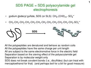
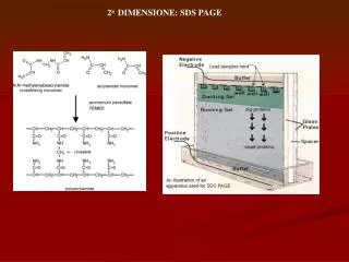
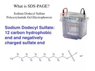
SDS-PAGE • Eindimensionale Auftrennungsmethode nach der Größe • DiskontinuirlichesTris-HCL / Tris-Glycin Puffersystem • Sammelgel: weitporig , pH 6,8 und geringere Pufferstärke sowie Stapelung der Proteine • Glycin: Folge-Ion wegen geringer Mobilität • Chlorid: Leit-Ion wegen hoher Mobilität SDS-PAGE Meike Flentje & Julia Schwab
SDS-PAGE • Trenngel: pH 8,8 sowie eng porig und damit erfahren die Proteine einen höheren Reibungswiderstand • Folge: bessere Auftrennung und schärfere Banden [3] SDS-PAGE Meike Flentje & Julia Schwab
SDS-PAGE Probenvorbereitung • Red. Laufpuffer mit einem Überschuss an SDS • Erwärmung der Proben auf 95 °C • Denaturierung • DTT spaltet die Disulfidbrücken • SDS kann sich anlagern und eine negative Mizelle bilden, damit werden Eigenladungen abgeschirmt • -> Gesamtladung ist proportional zur Proteingröße [4] SDS-PAGE Meike Flentje & Julia Schwab
Coomassie Gelfärbung • Farbmittel: Coomassie Brillant Blue R-250 • Unspezifische Proteinfärbung • Interaktion mit den basischen Resten, damit werden Lysin, Arginin und Histidin detektiert SDS-PAGE Meike Flentje & Julia Schwab
Versuchsdurchführung • Gel gießen • Probenvorbereitung • Proben auftragen • Gellauf • Coomassie Färbung SDS-PAGE Meike Flentje & Julia Schwab
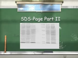
Ergebnisse Abb. 3: : 14: 34 h (A) (7 µL); 15-20: 34 h-44 h (B) (7 µL); 21: 46 h (A) (7 µL); M: Protein-Marker (3 µL); 22: Gesamtzellprotein (A) (7 µL); 23: lösliche Proteine (A) (7 µL); 24: unlösliche Proteine (A) (7 µL); 25: Gesamtzellprotein (B) (7 µL); 26: lösliche Proteine (B) (7 µL); 27: unlösliche Proteine (B) (7 µL) Abb. : SDS-PAGE nach der Kultivierung: 1: Vorkultur (A) (7 μL); 2-7: 12 h-22 h (B) (7 μL); 8-13: 22 h-32 h (A) (7 μL); M: Protein-Marker (3 μL) SDS-PAGE Meike Flentje & Julia Schwab
Ergebnisse Abb. 4: SDS-PAGE nach der Kationenaustausch-Membran-Chromatographie: Probe 1-23 (10 μL); M: Protein-Marker (3 μL) SDS-PAGE Meike Flentje & Julia Schwab
Ergebnisse Abb. 5: SDS-PAGE nach der Heparin-Sepharose-Affinitäts-Chromatographie: M: Protein-Marker (3 μL); 1-16: Probe (10 μL) SDS-PAGE Meike Flentje & Julia Schwab
Ergebnisse Abb. 6: Zusammenfassung der Ergebnisse: 1: Gesamtzellprotein (B) (10 μL); 2: lösliche Proteine (B) (10 μL); 3: unlösliche Proteine (B) (10 μL); M: Protein-Marker (4 μL); 4-5: Heparin-Sepharose-Affinitäts-Chromatographie K + M (15 μL) SDS-PAGE Meike Flentje & Julia Schwab
Vielen Dank für eure Aufmerksamkeit Fragen? SDS-PAGE Meike Flentje & Julia Schwab
Literatur • [1] Atherton, B. A.; Cunningham, E. L.; Splittgerber, A. G.: A mathematical model forthedescrip-tionoftheCoomassiebrilliantblueproteinassay. In: Anal Biochem. 233(2), S. 160-8 (1996). • [2] Fazekas de St. Groth, S.; Webster, R. G.; Datyner, A.: Twonewstainingproceduresforquantit-ativeestimationofproteins on electrophoreticstrips. In: Biochimica et Biophysica Acta. 71, S. 377–391 (1963). • [3] Laemmli, U. K.: Cleavageofstructuralproteinsduringtheassemblyoftheheadofbacterio-phage T4. In: Nature. 227, S. 680-685 (1970). • [4] Lottspeich, H.; Zorbas, H.: Bioanalytik. 1. Aufl. Heidelberg: Spektrum, Akad. Verl. (1998). • [5] Reinard, T.: Molekularbiologische Methoden. 1. Aufl. Stuttgart: Eugen Ulmer KG. (2010). SDS-PAGE Meike Flentje & Julia Schwab
Literatur - Bild • Bild 1: http://commons.wikimedia.org/wiki/File:Protein_FGF2_PDB_1bas.png; 28.05.2013 • Bild 2: http://www.pnas.org/content/96/7/3658/F2.expansion.html; 28.05.2013 • Bild 3: https://www.goldbio.com/FGF2-Basic-FGF-Human-P5983-C241.php; 28.05.2013 • Bild 4: http://www.bio-rad.com/evportal/en/US/LSR/Solutions/LUSOW4GRI/Protein-Electrophoresis-Methods; 28.05.2013 SDS-PAGE Meike Flentje & Julia Schwab