Download
1 / 59
590 likes | 710 Vues
Learn about pulmonary and systemic circuits, myocardial oxygen consumption, cardiac vs. skeletal muscle, arterial blood pressure, cardiac cycle, and factors influencing blood pressure. Explore heart rate regulation, stroke volume, and the impact of exercise on pulse pressure and mean arterial pressure. Gain insight into the Frank-Starling mechanism and factors influencing venous return.

E N D
Pulmonary and Systemic Circuits • Pulmonary circuit • Right side of the heart • Pumps deoxygenated blood to the lungs via pulmonary arteries • Returns oxygenated blood to the left side of the heart via pulmonary veins • Systemic circuit • Left side of the heart • Pumps oxygenated blood to the whole body via arteries • Returns deoxygenated blood to the right side of the heart via veins
Myocardium – “The Heart Wall” • Receives blood supply via coronary arteries • High demand for oxygen and nutrients even at rest • Myocardial Oxygen Consumption (MVO2) • Influenced by: • Force • Rate • Frequency • Rate Pressure Product (RPP): myocardial workload or stress • RPP = HR X SBP • Monitors exercise in patients with heart disease because value reflect cardiac function There is a linear correlation between RPP and MVO2
The Cardiac Cycle • Systole • Contraction phase • Ejection of blood • ~2/3 blood is ejected from ventricles per beat • Diastole • Relaxation phase • Filling with blood
Pressure Changes during the Cardiac Cycle • Diastole • Pressure in ventricles is low • Filling with blood from atria • AV valves open when ventricular P < atrial P • Systole • Pressure in ventricles rises when atria contract and rises even sharper when ventricles contract • Forces closure of AV valves to prevent backflow into atria • Blood ejected in pulmonary and systemic circulation • Semilunar valves open when ventricular P > aortic P • Heart sounds • First: closing of AV valves • Second: closing of aortic and pulmonary valves
Arterial Blood Pressure • Expressed as systolic/diastolic • Normal is 120/80 mmHg for males, 110/70 for females • Pulse pressure • Difference between systolic and diastolic • Represents force heart produces with each contraction • Indicator of mortality and risk for heart disease • Does not mean two individuals with equal pulse pressures have equal risk • Low = <25% of systolic mmHg value • Indicates poor left ventricular stroke volume or blood loss • High = stiffness of artery walls • Arterioscelrosisor atherosclerosis What happens to pulse pressure with exercise?
Mean Arterial Pressure (MAP) • Mean arterial pressure (MAP) • MAP = DBP + .33 (pulse pressure) • Determines rate of blood flow through systemic circuit • Average pressure in the arteries • Typically thought of as the “aortic pressure” • Can not be used during exercise, based on cardiac cycle at rest • Factors: • Cardiac output (Q) • Total vascular resistance • Clinical significance: perfusion pressure of organs in body • >60 mmHg is thought to be sufficient • 70-110 mmHg is normal
Factors that Influence Arterial Blood Pressure • Short-term regulation • Sympathetic nervous system • Baroreceptors in aorta and carotid arteries • Increase in BP = decreased SNS activity • Decrease in BP = increased SNS activity • Long-term regulation • Kidneys • Via control of blood volume • What hormone from kidney impacts blood pressure? • What was the name of the common medication?
Cardiac Output (Q) • The amount of blood pumped by the heart each minute • Product of heart rate and stroke volume • Heart rate • Number of beats per minute • = 208- 0.7(Age) • Stroke volume • Amount of blood ejected in each beat (mL) • Depends on: • Training state • Gender • Age • Disease Normal Levels: 5-6 L/min Exercise Levels: 20-35 L/min Q = HR x SV
Autonomic Nervous System Sympathetic Parasympathetic Ach Ach Ach NE Effects: * Increase HR * Increase contractility * Innervate Atria and Ventricles Effects: * Decrease HR * Decrease contractility * Innervate Atria
Stroke Volume (SV) • Regulation of SV (70 ml/beat) • End-diastolic volume (EDV) • Volume of blood in the ventricles at the end of diastole (“preload”) • Average aortic blood pressure • Pressure the heart must pump against to eject blood (“afterload”) • Mean arterial pressure • SV and afterload are inversely proportional • During exercise d/t arteriole dilation, afterload is minimized • Strength of the ventricular contraction (contractility) • Enhanced by: • Circulating epinephrine and norepinephrine • Direct sympathetic stimulation of heart • Stroke volume = EDV-ESV
End Diastolic Volume (EDV) – “Preload” • Frank-Starling mechanism • Greater EDV results in a more forceful contraction • Due to stretch of ventricles • Discuss! • Dependent on venous return • Venous return increased by: • Venoconstriction • Via SNS • Skeletal muscle pump • Rhythmic skeletal muscle contractions force blood in the extremities toward the heart • One-way valves in veins prevent backflow of blood • Respiratory pump • Changes in thoracic pressure pull blood toward heart
Video • Frank Starling Mechanism
Ejection Fraction (EF) • Percentage of blood pumped from left ventricle • Stroke Volume / EDV • 55-70% is considered normal • Helpful in diagnostics for: • Disease • Heart failure: systolic (HFrEF) or diastolic (HFpEF) • 40-55% indicates damage (heart attack or dilated cardiomyopathy) • >75% indicates hypertrophic cardiomyopathy • Efficiency • Function • Can be calculated via echocardiogram
Pressure Blood flow = Resistance Relationships Among Pressure, Resistance, and Flow • Blood flow • Directly proportional to the pressure difference between the two ends of the system • Inversely proportional to resistance • Pressure • Proportional to the difference between MAP and right atrial pressure ( Pressure) What causes resistance in the circulatory system?
Redistribution of Blood Flow During Exercise • Increased blood flow to working skeletal muscle • At rest, 15–20% of cardiac output to muscle • Increases to 80–85% during maximal exercise • Vasoconstriction to visceral organs and inactive tissues • Liver, kidneys, GI tract • Decreases to only 20-30% of resting values • SNS vasoconstriction • Redistribution depends on metabolic rate • Exercise intensity
Regulation of Local Blood Flow during Exercise • Skeletal muscle vasodilation • Autoregulation – what does this mean? • At beginning of exercise • Blood flow increased to meet metabolic demands of tissue • Due to changes in O2 tension, CO2 tension, nitric oxide, potassium, adenosine, and pH • Level of vasodilation is regulated by metabolic need of muscle
Cardiac Output Normal Values • Rest: 5-6 L/min • Exercise: 20-35 L/min
Heart Rate Normal Values • Rest: 50-70 beats/min • Exercise: 220-age (maximal)
Stroke Volume Normal Values • Rest: 70 ml/beat • Exercise: up to 200 ml/beat
Changes in Cardiac Output During Exercise • Cardiac output increases due to: • Increased HR • Linear increase to max • For adults: • For children: • Increased SV • Increase, then plateau at ~40% VO2 max in untrained • No plateau in highly trained subjects • WHY?! Max HR = 220 – age (years) Max HR = 208 – 0.7 x age (years)
Arteriovenous Oxygen Difference(a-vO2diff) • Difference in oxygen content of blood from arteries to veins • Amount of O2 that is taken up from 100 ml blood • Calculated in two ways: • Blood samples • Fick Equation • VO2= HR x SV x A-VO2diff
Changes in Arterial-Mixed Venous O2 Content During Single Bout of Exercise • Higher arteriovenous difference (a-vO2 difference) • Increase due to higher amount of O2 taken up • Used for oxidative ATP production • Factors • Extraction at muscles • Delivery of oxygen rich blood At the onset of exercise, do you think that A-vO2 is noticed right away? Why/why not?
Circulatory Responses to Single Bout of Exercise • Transition from Rest to Exercise • Rapid increase in HR, SV, and Q • If work rate constant and below lactate threshold • Steady state plateau in HR, SV, and Q within 2-3 minutes • Recovery from Exercise • Rapid from short-term, low intensity exercise • Recovery speeds varies: Trained vs. untrained • Slower with long-term exercise • Particularly in hot/humid environments
Incremental Exercise • Heart rate and cardiac output • Increases linearly with increasing work rate • Reaches plateau at 100% VO2 max • Blood pressure • Mean arterial pressure (MAP) increases linearly • Systolic BP increases • Diastolic BP remains fairly constant • Rate pressure product • Increases linearly with exercise intensity • Exercise v. rest: 5x • Used to prescribe exercise to cardiac patients
Intermittent Exercise • Recovery of heart rate and blood pressure between bouts depend on: • Fitness level • Temperature and humidity • Duration and intensity of exercise
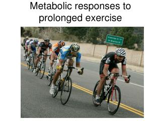
Prolonged Exercise • Cardiac output is maintained • Gradual decrease in stroke volume • Due to dehydration and reduced plasma volume • Gradual increase in heart rate • Cardiovascular drift • Helps keep constant Q
VO2Max • Maximal oxygen uptake • Most valid measurement of cardiovascular fitness • “Critical point during exhaustive exercise in which oxygen uptake reaches a plateau, beyond which greater increments in exercise intensity elicit no further rise in oxygen uptake.” • Physiological ceiling – what do I mean by this again?
VO2max • Affected by genetics and training, age, gender… • Dependent on: • Maximum ability of cardiorespiratory system to deliver oxygen to the muscle • Ability of muscles to use oxygen and produce ATP aerobically
VO2max Test Termination Criteria Exhaustion 4 • Plateau in O2 uptake with • increasing intensity • ( of < 2.1 ml/kg/min) RER VO2 (L/min) 2. Reach RER of > 1.1 1.1 3 8 mmol • Reach blood lactate of • > 8 mmol 0.8 Lactate • Reach age-predicted Hrmax • (220 – age) 2 mmol 2 0 5 10 15 % Grade
VO2 Max Can VO2max be Calculated? *Fick Equation * VO2max = HRmax x SVmax x A-VO2diffmax = QmaxX A-VO2diffmax Example: Q: What is the VO2max of a person with a HRmaxof 185 b/min, SVmaxof 150 ml/b, and A-VO2diff of 15 ml/100 ml blood (assume all are max values)? A: 4.16 L/min
VO2max and Genetics Genetic (untrained) VO2max (ml/kg/min)Population 551 in 6 people 651 in 40 people 701 in 2,000 people 80“Very, very Few” From: Shephard and Astrand, 1992
Gender and Aging 5 Men typically have a ~15% higher VO2max (ml/kg/min) than women VO2max declines ~ 1% each year after age 20 VO2 (L/min) 3 M F 1 10 20 30 40 50 60 Age
VO2max and Gender Q: Why do men typically have higher VO2max values than women? A: (1) More lean body mass (active tissue that can use O2) Example: Man weighs 70 kg and has VO2max of 4.5L/min = 64.3 ml/kg/min Woman weighs 60 kg and has VO2max of 3.3L/min = 55.0 ml/kg/min = 14.5% higher in man If man is 15% body fat (59.5 kg is FFM), then VO2max = ~ 75.6 ml/kgFFM/min If woman is 22% body fat (47 kg is FFM), then VO2max = ~ 70.5 ml/kgFFM/min = 6.7% higher in man Why is VO2max still higher in man after controlling for LBM?
VO2max and Gender Q: Why do men typically have higher VO2max values than women? A: (2) Higher Hb concentration (Larger O2 carrying capacity) • Carry more oxygen • Transport more oxygen
Oxygen Transport: Limitations to VO2max Hemoglobin Ventilation Cardiac Output Muscle Mass/ Circulation/ Metabolism