Download
1 / 37
370 likes | 534 Vues
The Nervous System: Structure and Control of Movement Dr. Kyle Coffey. Week 6&7. The Nervous System. Way the body perceives, adapts, and responds to the internal and external environments Touch, pain, temperature 4 major functions:

E N D
The Nervous System:Structure and Control of MovementDr. Kyle Coffey Week 6&7
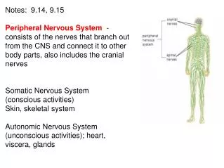
The Nervous System • Way the body perceives, adapts, and responds to the internal and external environments • Touch, pain, temperature • 4 major functions: • Control of the internal environment (with what other system?) • Voluntary control of movement • Spinal cord reflexes • Assimilation of experiences necessary for memory and learning
Electrical Activity in Neurons • Neurons are an “excitable tissue” due to: • Irritability • Ability to respond to a stimulus and convert it to a neural impulse • Conductivity • Transmission of the impulse along the axon • Electrical signal is initiated by a stimulus that causes a change in the electrical charge of the neuron
Resting Membrane Potential (RMP) • Neuron is polarized • Negative charge inside cells at rest compared to outside creates electrical charge difference • -5 to -100 mv in all other cells • -40 to -75 mv in neurons • Determined by: • Permeability of plasma membrane to ions • Difference in ion concentrations across membrane • Na+, K+, Cl–, and Ca++
Resting Membrane Potential (RMP) • Maintained by sodium-potassium pump • Proteins that act as channels with gates that open and close • Use ATP to maintain RMP • 2/3 of cell’s energy expenditure • Na+/K+ pump moves 2 K+ in and 3 Na+ out • At rest, almost all Na+ channels are closed and few K+ channels open • “Leak” of K+ out of cell versus Na+ in • Overall, Results in net loss of (+) charges out of cell, resulting in (-) RMP
Action Potential • Depolarization: Occurs when a stimulus of sufficient strength causes inside of cell to become more positive • Critical threshold • Opens Na+channels and Na+ diffuses into cell • Repolarization • Return to resting membrane potential • K+ leaves the cell rapidly • Na+ channels close • Inside becomes more negative until RMP is reached • All-or-none law • Once a nerve impulse is initiated, it will travel the length of the neuron
Synaptic Transmission and Neurotransmitters • Synapse • Small gap (20-30 nM)between presynaptic neuron and postsynaptic neuron • Synaptic Transmission • Occur when sufficient amounts of a specific transmitter are released from synaptic vesicles in presynaptic neuron • Neurotransmitter • Chemical messenger released from presynaptic membrane • Binds to receptor on postsynaptic membrane • Causes depolarization of postsynaptic membrane (excitatory)
Video • RMP & Action Potential
Summation • Temporal summation • Summing several EPSPs from one presynaptic neuron • Rapid, repetitive excitation • Spatial summation • Summing from several different presynaptic neurons
Acetylcholine • Transmitter of the nerve/muscle junction • Binds to receptors on post-synaptic neuron • Opens channels that permit Na+ to enter nerve or muscle cell • Broken down by enzymes in synaptic cleft to prevent chronic depolarization • Acetylcholinesterase • Can be both excitatory and inhibitory • Depolarization of skeletal muscle • Re/hyperpolarization of heart (slows HR)
Joint Proprioceptors • Free nerve endings • Most abundant • Sensitive to touch and pressure • Initially strongly stimulated at beginning of movement, then adapt • Golgi-type receptors • Not as abundant • Found in ligaments and around joints • Similar to free nerve endings • Pacinian corpuscles • In tissues around joints • Adapt rapidly following the initiation of movement • Detect rate of joint rotation
Sensory Information and Reflexes • Proprioceptors • Receptors that provide CNS with information about body position • Located in joints and muscles • Muscle spindles, Golgi tendon organs, joint receptors • Kinesthesia • Conscious recognition of the position of body parts • Limb movement rates
Muscle Proprioceptors • Provide sensory feedback to nervous system • Tension development by muscle • Account of muscle length • Chemoreceptors • Send info to CNS about: • Muscle pH • Concentrations of extracellular potassium • Changes in O2 + CO2 • Provide CNS with information about metabolic rate of muscular activity • Important in regulation of cardiovascular and pulmonary responses • Muscle spindle • Golgi tendon organ
Muscle Spindle • Found in large numbers • Responds to changes in muscle length • Consists of: • Intrafusal fibers (thin muscle fibers) surrounded by CT sheath • Run parallel to normal muscle fibers (extrafusal fibers) • Sensory nerve endings • Primary: dymanic changes in length • Secondary: static muscle length • Gamma motor neurons • Stimulate intrafusal fibers to contract simultaneously with extrafusal fibers (by alpha motor neuron)
Video • Muscle Spindle
Golgi Tendon Organs (GTOs) • Located within tendon • In series (parallel) with extrafusal fibers • Monitors tension developed in muscle • “Safety devices” that prevent muscle damage during excessive force generation • Stimulation results in reflex relaxation of muscle • Ability to voluntarily oppose GTO inhibition may be related to gains in strength • Also responsible for inverse stretch reflex (inverse myotactic) • Decreased ms tension with vigorous contraction
Video • Golgi Tendon Organs (GTO)
Types of Training or Activity • Plyometric • Strength Training • Cross-Fit • Running • Biking • Stretching
Withdrawal Reflex • Reflex contraction of skeletal muscle • Occurs in response to sensory input • Not dependent on higher brain centers (spinal level ONLY) • Reciprocal inhibition • EPSPs to muscles to withdraw from stimulus • IPSPs to antagonistic muscles • Crossed-extensor reflex • Opposite limb supports body during withdrawal of injured limb
Motor Unit Recruitment and the Size Principle • Motor unit recruitment • Recruitment of more muscle fibers through motor unit activation • Orderly fashion and as a function of their size • The size principle • Smallest motor units recruited first • Produce action potential sooner • Types of motor units • Type S (slow) or type I fibers [smallest] • Type FR (fast, fatigue resistant) or type IIa fibers [intermediate] • Type FF (fast, fatigable) or type IIb/x fibers [largest] • Recruitment pattern during incremental exercise • Type S (1) type FR (IIa) type FF (IIb)
Motor Functions of Spinal Cord • Withdrawal reflex • Other reflexes • Important for the control of voluntary movement • Spinal tuning • Voluntary movement translated into appropriate muscle action • Higher brain centers concerned with general parameters of movement • Details of movement refined in spinal cord
Autonomic Nervous System • Operates below conscious level, closely linked to emotion • Responsible for maintaining internal environment • Effector organs not under voluntary control • Smooth muscle, cardiac muscle, and glands • Sympathetic division • Releases norepinephrine (NE) • Excites an effector organ • After stimulation, NE is removed from synapse or inactivated • Parasympathetic division • Releases acetylcholine (ACh) • Inhibits effector organ • After stimulation, ACh is degraded by acetylcholinesterase