Download
1 / 173
1.79k likes | 2.11k Vues
16 Neural Integration II: The Autonomic Nervous System and Higher-Order Functions. An Introduction to the ANS and Higher-Order Functions. Learning Outcomes 16-1 Compare the organization of the autonomic nervous system with that of the somatic nervous system.

E N D
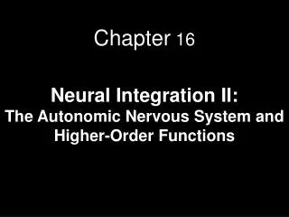
16 Neural Integration II: The Autonomic Nervous System and Higher-Order Functions
An Introduction to the ANS and Higher-Order Functions Learning Outcomes 16-1 Compare the organization of the autonomic nervous system with that of the somatic nervous system. 16-2 Describe the structures and functions of the sympathetic division of the autonomic nervous system. 16-3 Describe the mechanisms of sympathetic neurotransmitter release and their effects on target organs and tissues.
An Introduction to the ANS and Higher-Order Functions Learning Outcomes 16-4 Describe the structures and functions of the parasympathetic division of the autonomic nervous system. 16-5 Describe the mechanisms of parasympathetic neurotransmitter release and their effects on target organs and tissues. 16-6 Discuss the functional significance of dual innervation and autonomic tone. 16-7 Describe the hierarchy of interacting levels of control in the autonomic nervous system, including the significance of visceral reflexes.
An Introduction to the ANS and Higher-Order Functions Learning Outcomes 16-8 Explain how memories are created, stored, and recalled, and distinguish among the levels of consciousness and unconsciousness. 16-9 Describe some of the ways in which the interactions of neurotransmitters influence brain function. 16-10 Summarize the effects of aging on the nervous system and give examples of interactions between the nervous system and other organ systems.
An Introduction to the ANS and Higher-Order Functions • Somatic Nervous System (SNS) • Operates under conscious control • Seldom affects long-term survival • SNS controls skeletal muscles • Autonomic Nervous System (ANS) • Operates without conscious instruction • ANS controls visceral effectors • Coordinates system functions • Cardiovascular, respiratory, digestive, urinary, reproductive
Figure 16-1 An Overview of Neural Integration OVERVIEW OF NEURAL INTEGRATION CHAPTER 16 CHAPTER 15 Sensoryprocessingcenters inbrain Higher-Order Functions Conscious andsubconsciousmotor centersin brain Memory, learning, andintelligence mayinfluence interpretationof sensory informationand nature of motoractivities Motorpathways Sensorypathways SomaticNervousSystem (SNS) AutonomicNervousSystem (ANS) Visceral effectors(examples: smoothmuscle, glands,cardiac muscle,adipocytes) Generalsensoryreceptors Skeletalmuscles
16-1 Autonomic Nervous System • Organization of the ANS • Integrative centers • For autonomic activity in hypothalamus • Neurons comparable to upper motor neurons in SNS
16-1 Autonomic Nervous System • Organization of the ANS • Visceral motor neurons • In brain stem and spinal cord, are known as preganglionic neurons • Preganglionic fibers • Axons of preganglionic neurons • Leave CNS and synapse on ganglionic neurons
16-1 Autonomic Nervous System • Visceral Motor Neurons • Autonomic ganglia • Contain many ganglionic neurons • Ganglionic neurons innervate visceral effectors • Such as cardiac muscle, smooth muscle, glands, and adipose tissue • Postganglionic fibers • Axons of ganglionic neurons
Figure 16-2a The Organization of the Somatic and Autonomic Nervous Systems Upper motorneurons inprimary motorcortex BRAIN Somatic motornuclei of brainstem SPINALCORD Lowermotorneurons Skeletalmuscle Somaticmotor nucleiof spinal cord Skeletalmuscle Somatic nervous system
Figure 16-2b The Organization of the Somatic and Autonomic Nervous Systems Visceral motornuclei inhypothalamus BRAIN Preganglionicneuron Visceral Effectors Smoothmuscle Autonomicnuclei inbrain stem Autonomicganglia Glands Cardiacmuscle Ganglionicneurons SPINALCORD Adipocytes Autonomicnuclei inspinal cord Preganglionicneurons Autonomic nervous system
16-1 Divisions of the ANS • The Autonomic Nervous System • Operates largely outside our awareness • Has two divisions • Sympathetic division • Increases alertness, metabolic rate, and muscular abilities • Parasympathetic division • Reduces metabolic rate and promotes digestion
16-1 Divisions of the ANS • Sympathetic Division • “Kicks in” only during exertion, stress, or emergency • “Fight or flight” • Parasympathetic Division • Controls during resting conditions • “Rest and digest”
16-1 Divisions of the ANS • Sympathetic and Parasympathetic Division • Most often, these two divisions have opposing effects • If the sympathetic division causes excitation, the parasympathetic causes inhibition • The two divisions may also work independently • Only one division innervates some structures • The two divisions may work together, with each controlling one stage of a complex process
16-1 Divisions of the ANS • Sympathetic Division • Preganglionic fibers (thoracic and superior lumbar; thoracolumbar) synapse in ganglia near spinal cord • Preganglionic fibers are short • Postganglionic fibers are long • Prepares body for crisis, producing a “fight or flight” response • Stimulates tissue metabolism • Increases alertness
16-1 Divisions of the ANS • Seven Responses to Increased Sympathetic Activity • Heightened mental alertness • Increased metabolic rate • Reduced digestive and urinary functions • Energy reserves activated • Increased respiratory rate and respiratory passageways dilate • Increased heart rate and blood pressure • Sweat glands activated
16-1 Divisions of the ANS • Parasympathetic Division • Preganglionic fibers originate in brain stem and sacral segments of spinal cord; craniosacral • Synapse in ganglia close to (or within) target organs • Preganglionic fibers are long • Postganglionic fibers are short • Parasympathetic division stimulates visceral activity • Conserves energy and promotes sedentary activities
16-1 Divisions of the ANS • Five Responses to Increased Parasympathetic Activity • Decreased metabolic rate • Decreased heart rate and blood pressure • Increased secretion by salivary and digestive glands • Increased motility and blood flow in digestive tract • Urination and defecation stimulation
16-1 Divisions of the ANS • Enteric Nervous System (ENS) • Third division of ANS • Extensive network in digestive tract walls • Complex visceral reflexes coordinated locally • Roughly 100 million neurons • All neurotransmitters are found in the brain
16-2 The Sympathetic Division • The Sympathetic Division • Preganglionic neurons located between segments T1 and L2 of spinal cord • Ganglionic neurons in ganglia near vertebral column • Cell bodies of preganglionic neurons in lateral gray horns • Axons enter ventral roots of segments
Figure 16-3 The Organization of the Sympathetic Division of the ANS Sympathetic Division of ANS Target Organs Ganglionic Neurons PreganglionicNeurons Visceral effectorsin thoracic cavity,head, body wall,and limbs Sympatheticchain ganglia(paired) Lateral grayhorns of spinalsegmentsT1–L2 Collateralganglia(unpaired) Visceral effectorsin abdominopelviccavity Adrenalmedullae(paired) Organs and systemsthroughout body KEY Preganglionic fibers Postganglionic fibers Hormones releasedinto circulation
16-2 The Sympathetic Division • Ganglionic Neurons • Occur in three locations • Sympathetic chain ganglia • Collateral ganglia • Suprarenal medullae
16-2 The Sympathetic Division • Sympathetic Chain Ganglia • Are on both sides of vertebral column • Control effectors: • In body wall • Inside thoracic cavity • In head • In limbs
Figure 16-4a Sites of Ganglia in Sympathetic Pathways SYMPATHETIC CHAIN GANGLIA Autonomic ganglion ofright sympathetic chain Preganglionicneuron Spinal nerve Autonomic ganglionof left sympathetic chain Innervatesvisceraleffectors viaspinal nerves White ramus Sympathetic nerve(postganglionicfibers) Ganglionicneuron Gray ramus Innervates visceralorgans in thoraciccavity viasympathetic nerves KEY Preganglionic neurons Ganglionic neurons
16-2 The Sympathetic Division • Collateral Ganglia • Are anterior to vertebral bodies • Contain ganglionic neurons that innervate tissues and organs in abdominopelvic cavity
Figure 16-4b Sites of Ganglia in Sympathetic Pathways COLLATERAL GANGLIA Lateralgrayhorn Whiteramus Splanchnicnerve(preganglionicfibers) Collateralganglion Innervatesvisceral organs inabdominopelviccavity Postganglionicfibers KEY Preganglionic neurons Ganglionic neurons
16-2 The Sympathetic Division • Adrenal Medullae (SuprarenalMedullae) • Very short axons • When stimulated, release neurotransmitters into bloodstream (not at synapse) • Function as hormones to affect target cells throughout body
Figure 16-4c Sites of Ganglia in Sympathetic Pathways THE ADRENAL MEDULLAE Preganglionic fibers Adrenalmedullae Secretesneurotransmittersinto generalcirculation Endocrine cells(specialized ganglionicneurons) KEY Preganglionic neurons Ganglionic neurons
16-2 The Sympathetic Division • Fibers in Sympathetic Division • Preganglionic fibers • Are relatively short • Ganglia located near spinal cord • Postganglionic fibers • Are relatively long, except at adrenal medullae
16-2 The Sympathetic Division • Organization and Anatomy of the Sympathetic Division • Ventral roots of spinal segments T1–L2 contain sympathetic preganglionic fibers • Give rise to myelinated white ramus • Carry myelinated preganglionic fibers into sympathetic chain ganglion • May synapse at collateral ganglia or in adrenal medullae
16-2 The Sympathetic Division • Sympathetic Chain Ganglia • Preganglionic fibers • One preganglionic fiber synapses on many ganglionic neurons • Fibers interconnect sympathetic chain ganglia • Each ganglion innervates particular body segment(s)
16-2 The Sympathetic Division • Sympathetic Chain Ganglia • Postganglionic Fibers • Paths of unmyelinated postganglionic fibers depend on targets
16-2 The Sympathetic Division • Sympathetic Chain Ganglia • Postganglionic fibers control visceral effectors • In body wall, head, neck, or limbs • Enter gray ramus • Return to spinal nerve for distribution • Postganglionic fibers innervate effectors • Sweat glands of skin • Smooth muscles in superficial blood vessels
16-2 The Sympathetic Division • Sympathetic Chain Ganglia • Postganglionic fibers innervating structures in thoracic cavity form bundles • Sympathetic nerves
16-2 The Sympathetic Division • Sympathetic Chain Ganglia • Each sympathetic chain ganglia contains: • 3 cervical ganglia • 10–12 thoracic ganglia • 4–5 lumbar ganglia • 4–5 sacral ganglia • 1 coccygeal ganglion
16-2 The Sympathetic Division • Sympathetic Chain Ganglia • Preganglionic neurons • Limited to spinal cord segments T1–L2 • White rami (myelinated preganglionic fibers) • Innervate neurons in: • Cervical, inferior lumbar, and sacral sympathetic chain ganglia
16-2 The Sympathetic Division • Sympathetic Chain Ganglia • Chain ganglia provide postganglionic fibers • Through gray rami (unmyelinated postganglionic fibers) • To cervical, lumbar, and sacral spinal nerves
16-2 The Sympathetic Division • Sympathetic Chain Ganglia • Only spinal nerves T1–L2 have white rami • Every spinal nerve has gray ramus • That carries sympathetic postganglionic fibers for distribution in body wall
16-2 The Sympathetic Division • Sympathetic Chain Ganglia • Postganglionic sympathetic fibers • In head and neck leave superior cervical sympathetic ganglia • Supply the regions and structures innervated by cranial nerves III, VII, IX, X
Figure 16-5 The Distribution of Sympathetic Innervation PONS Superior Middle Cervicalsympatheticganglia Inferior T1 T1 Gray rami tospinal nerves KEY Preganglionic neurons Ganglionic neurons
Figure 16-5 The Distribution of Sympathetic Innervation Eye PONS Salivaryglands Sympathetic nerves Heart Cardiac andpulmonary plexuses(see Figure 16-10) T1 Lung KEY Preganglionic neurons Ganglionic neurons
Figure 16-5 The Distribution of Sympathetic Innervation T1 Greatersplanchnicnerve KEY Preganglionic neurons Ganglionic neurons Celiac ganglion Superiormesentericganglion Liver and gallbladder Stomach Splanchnicnerves Spleen Pancreas Largeintestine Smallintestine Inferiormesentericganglion L2 Adrenalmedulla Kidney Coccygealganglia (Co1)fused together Uterus Penis Urinary bladder Scrotum Ovary
Figure 16-5 The Distribution of Sympathetic Innervation Postganglionic fibersto spinal nerves(innervating skin,blood vessels,sweat glands,arrector pili muscles,adipose tissue) L2 Sympatheticchain ganglia Spinal cord KEY Preganglionic neurons Ganglionic neurons
16-2 The Sympathetic Division • Collateral Ganglia • Receive sympathetic innervation via sympathetic preganglionic fibers • Splanchnic nerves • Formed by preganglionic fibers that innervate collateral ganglia • In dorsal wall of abdominal cavity • Originate as paired ganglia (left and right) • Usually fuse together in adults
16-2 The Sympathetic Division • Collateral Ganglia • Postganglionic fibers • Leave collateral ganglia • Extend throughout abdominopelvic cavity • Innervate variety of visceral tissues and organs • Reduction of blood flow and energy by organs not vital to short-term survival • Release of stored energy reserves
16-2 The Sympathetic Division • Collateral Ganglia • Preganglionic fibers from seven inferior thoracic segments • End at celiac ganglion or superior mesenteric ganglion • Ganglia embedded in network of autonomic nerves • Preganglionic fibers from lumbar segments • Form splanchnic nerves • End at inferior mesenteric ganglion
16-2 The Sympathetic Division • Collateral Ganglia • Celiac ganglion • Pair of interconnected masses of gray matter • May form single mass or many interwoven masses • Postganglionic fibers innervate stomach, liver, gallbladder, pancreas, and spleen
16-2 The Sympathetic Division • Collateral Ganglia • Superior mesenteric ganglion • Near base of superior mesenteric artery • Postganglionic fibers innervate small intestine and proximal 2/3 of large intestine
16-2 The Sympathetic Division • Collateral Ganglia • Inferior mesenteric ganglion • Near base of inferior mesenteric artery • Postganglionic fibers provide sympathetic innervation to portions of: • Large intestine • Kidney • Urinary bladder • Sex organs
16-2 The Sympathetic Division • Adrenal Medullae • Preganglionic fibers entering adrenal gland proceed to center (adrenal medulla) • Modified sympathetic ganglion • Preganglionic fibers synapse on neuroendocrine cells • Specialized neurons secrete hormones into bloodstream