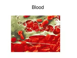
Blood and its Components
690 likes | 713 Vues
This article provides an overview of blood and its components, including the physical characteristics of blood, the composition of blood plasma, and the different formed elements such as erythrocytes, leukocytes, and platelets. It also discusses the functions and abnormalities of these components.

Blood and its Components
E N D
Presentation Transcript
10 Blood
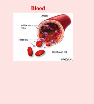
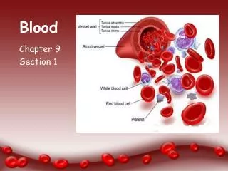
Blood • The only fluid tissue in the human body • Classified as a connective tissue • Components of blood • Living cells • Formed elements • Non-living matrix • Plasma
Blood • If blood is centrifuged • Red blood cells sink to the bottom (45 percent of blood, a percentage known as the hematocrit) • Buffy coat is a thin, whitish layer between the erythrocytes and plasma • Plasma rises to the top (55 percent of blood) • Everything else (less than 1 percent of blood)
Physical Characteristics of Blood • Color range • Oxygen-rich blood is scarlet (bright) red • Oxygen-poor blood is dull red • pH must remain between 7.35–7.45 • Blood temperature is slightly higher than body temperature at 100.4°F • In a healthy man, blood volume is about 5–6 liters • Blood makes up 8 percent of body weight
Blood Plasma • Composed of approximately 90 percent water • Includes many dissolved substances • Nutrients • Salts (electrolytes) • Respiratory gases • Hormones • Plasma proteins • Waste products
Blood Plasma • Plasma proteins • Most abundant solutes in plasma • Most plasma proteins are made by liver • Various plasma proteins include • Albumin—regulates osmotic pressure • Clotting proteins—help to stem blood loss when a blood vessel is injured • Antibodies—help protect the body from pathogens
Blood Plasma (skip) • Acidosis • Blood becomes too acidic • Alkalosis • Blood becomes too basic • In each scenario, the respiratory system and kidneys help restore blood pH to normal
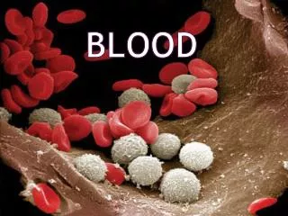
Formed Elements • Erythrocytes • Red blood cells (RBCs) • Leukocytes • White blood cells (WBCs) • Platelets • Cell fragments
Lymphocyte Platelets Erythrocytes Neutrophils Figure 10.2
Formed Elements • Erythrocytes (red blood cells or RBCs) • Main function is to carry oxygen • Anatomy of circulating erythrocytes • Biconcave disks • Essentially bags of hemoglobin • Anucleate (no nucleus) • Contain very few organelles • 5 million RBCs per cubic millimeter of blood
Formed Elements • Hemoglobin • Iron-containing protein • Binds strongly, but reversibly, to oxygen • Each hemoglobin molecule has four oxygen binding sites • Each red blood cell has 250 million hemoglobin molecules • Normal blood contains 12–18 g of hemoglobin per 100 mL blood
Formed Elements (skip) • Homeostatic imbalance of RBCs • Anemia is a decrease in the oxygen-carrying ability of the blood • Sickle cell anemia (SCA) results from abnormally shaped hemoglobin • Polycythemia is an excessive or abnormal increase in the number of erythrocytes
Formed Elements (Skip) • Polcythemia • Disorder resulting from excessive or abnormal increase of RBC • May be caused by bone marrow cancer (polycythemia vera) • May be a response to life at higher altitudes (secondary polycythemia) • Increased RBC slows blood flow and increases blood viscosity
Formed Elements • Leukocytes (white blood cells or WBCs) • Crucial in the body’s defence against disease • These are complete cells, with a nucleus and organelles • Able to move into and out of blood vessels (diapedesis) • Can move by ameboid motion • Can respond to chemicals released by damaged tissues • 4,800 to 10,800 WBC per cubic millimeter of blood
Formed Elements (skip) • Abnormal numbers of leukocytes • Leukocytosis • WBC count above 11,000 leukocytes/mm3 • Generally indicates an infection • Leukopenia • Abnormally low leukocyte level • Commonly caused by certain drugs such as corticosteroids and anticancer agents • Leukemia • Bone marrow becomes cancerous, turns out excess WBC
Formed Elements (skip) • Types of leukocytes • Granulocytes • Granules in their cytoplasm can be stained • Possess lobed nuclei • Include neutrophils, eosinophils, and basophils • Agranulocytes • Lack visible cytoplasmic granules • Nuclei are spherical, oval, or kidney-shaped • Include lymphocytes and monocytes
Easy way to remember this list Never Let Monkeys Eat Bananas Formed Elements (skip) • List of the WBCs from most to least abundant • Neutrophils • Lymphocytes • Monocytes • Eosinophils • Basophils
Formed Elements (skip) • Types of granulocytes • Neutrophils • Cytoplasm stains pale pink and contains fine granules • Deep purple nucleus contains three to seven lobes • Function as phagocytes at active sites of infection • Numbers increase during infection • 3,000–7,000 neutrophils in a cubic millimeter of blood (40–70% of WBCs)
Formed Elements (skip) • Types of granulocytes (continued) • Eosinophils • Red, coarse cytoplasmic granules • Figure-8 or bilobed nucleus stains blue-red • Function to kill parasitic worms and play a role in allergy attacks • 100–400 eosinophils in a cubic millimeter of blood (1–4% of WBCs)
Formed Elements (Skip) • Types of granulocytes (continued) • Basophils • Sparse but large blue-purple granules • U- or S-shaped nucleus stains dark blue • Release histamine (vasodilator) at sites of inflammation • Contain heparin (anticoagulant) • 20–50 basophils in a cubic millimeter of blood (0–1% of WBCs)
Formed Elements (skip) • Types of agranulocytes • Lymphocytes • Cytoplasm is pale blue • Dark purple-blue nucleus • Functions as part of the immune response • B lymphocytes produce antibodies • T lymphocytes are involved in graft rejection, fighting tumors and viruses • 1,500–3,000 lymphocytes in a cubic millimeter of blood (20–45% of WBCs)
Formed Elements (skip) • Types of agranulocytes (continued) • Monocytes • Largest of the white blood cells • Gray-blue cytoplasm • Dark blue-purple nucleus is often kidney shaped • Function as macrophages • Important in fighting chronic infection • 100–700 monocytes per cubic millimeter of blood (4–8% of WBCs)
Formed Elements • Platelets • Derived from ruptured cells multinucleate cells (megakaryocytes) • Needed for the clotting process • Platelet count ranges from 150,000 to 400,000 per cubic millimeter of blood • 300,000 is considered a normal number of platelets per cubic millimeter of blood
Hemostasis • Platelet plug formation • Collagen fibers are exposed by a break in a blood vessel • Platelets become “sticky” and cling to fibers • Anchored platelets release chemicals to attract more platelets • Platelets pile up to form a platelet plug
Hematopoiesis • Blood cell formation • Occurs in red bone marrow • All blood cells are derived from a common stem cell (hemocytoblast) • Hemocytoblast differentiation • Lymphoid stem cell produces lymphocytes • Myeloid stem cell produces all other formed elements
Formation of Erythrocytes • Unable to divide, grow, or synthesize proteins • Wear out in 100 to 120 days • When worn out, RBCs are eliminated by phagocytes in the spleen or liver • Lost cells are replaced by division of hemocytoblasts in the red bone marrow
Control of Erythrocyte Production • Rate is controlled by a hormone (erythropoietin) • Kidneys produce most erythropoietin as a response to reduced oxygen levels in the blood • Homeostasis is maintained by negative feedback from blood oxygen levels
IMBALANCE Homeostasis: Normal blood oxygen levels 1 Stimulus Low blood O2−carrying ability due to • Decreased RBC count • Decreased amount of hemoglobin • Decreased availability of O2 5 O2−carrying ability of blood increases. IMBALANCE 4 Enhanced erythropoiesis increases RBC count. 2 Kidney (and liver to a smaller extent) releases erythropoietin 3 Erythropoietin stimulates red bone marrow. Figure 10.5
IMBALANCE Homeostasis: Normal blood oxygen levels 1 Stimulus Low blood O2−carrying ability due to • Decreased RBC count • Decreased amount of hemoglobin • Decreased availability of O2 IMBALANCE Figure 10.5, step 1
IMBALANCE Homeostasis: Normal blood oxygen levels 1 Stimulus Low blood O2−carrying ability due to • Decreased RBC count • Decreased amount of hemoglobin • Decreased availability of O2 IMBALANCE 2 Kidney (and liver to a smaller extent) releases erythropoietin Figure 10.5, step 2
IMBALANCE Homeostasis: Normal blood oxygen levels 1 Stimulus Low blood O2−carrying ability due to • Decreased RBC count • Decreased amount of hemoglobin • Decreased availability of O2 IMBALANCE 2 Kidney (and liver to a smaller extent) releases erythropoietin 3 Erythropoietin stimulates red bone marrow. Figure 10.5, step 3
IMBALANCE Homeostasis: Normal blood oxygen levels 1 Stimulus Low blood O2−carrying ability due to • Decreased RBC count • Decreased amount of hemoglobin • Decreased availability of O2 IMBALANCE 4 Enhanced erythropoiesis increases RBC count. 2 Kidney (and liver to a smaller extent) releases erythropoietin 3 Erythropoietin stimulates red bone marrow. Figure 10.5, step 4
IMBALANCE Homeostasis: Normal blood oxygen levels 1 Stimulus Low blood O2−carrying ability due to • Decreased RBC count • Decreased amount of hemoglobin • Decreased availability of O2 5 O2−carrying ability of blood increases. IMBALANCE 4 Enhanced erythropoiesis increases RBC count. 2 Kidney (and liver to a smaller extent) releases erythropoietin 3 Erythropoietin stimulates red bone marrow. Figure 10.5, step 5
Formed Elements (Skip) • Polycythemia • Disorder resulting from excessive or abnormal increase of RBC • May be caused by bone marrow cancer (polycythemia vera) • May be a response to life at higher altitudes (secondary polycythemia) • Increased RBC slows blood flow and increases blood viscosity
Formation of White Blood Cells and Platelets • Controlled by hormones • Colony stimulating factors (CSFs) and interleukins prompt bone marrow to generate leukocytes • Thrombopoietin stimulates production of platelets
Hemostasis • Stoppage of bleeding resulting from a break in a blood vessel • Hemostasis involves three phases • Vascular spasms • Platelet plug formation • Coagulation (blood clotting)
Hemostasis • Vascular spasms • Vasoconstriction of smooth muscle causes blood vessel to spasm • Spasms narrow the blood vessel, decreasing blood loss
1 Step Vascular spasms occur. • Smooth muscle contracts, causing vasoconstriction. Figure 10.6, step 1
Hemostasis • Platelet plug formation • Collagen fibers are exposed by a break in a blood vessel • Platelets become “sticky” and cling to fibers • Anchored platelets release chemicals to attract more platelets • Platelets pile up to form a platelet plug
2 Step Platelet plug forms. • Injury to lining of vessel exposes collagen fibers; • platelets adhere. Collagen fibers • Platelets release chemicals that make nearby • platelets sticky; platelet plug forms. Platelets Figure 10.6, step 2
Hemostasis • Coagulation • Injured tissues release tissue factor (TF) • PF3 (a phospholipid) interacts with TF, blood protein clotting factors, and calcium ions to trigger a clotting cascade • Prothrombin activator converts prothrombin to thrombin (an enzyme)