Understanding the Nervous System: Structure, Function, and Neurons
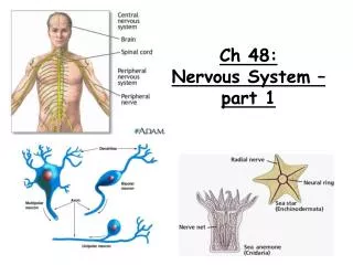
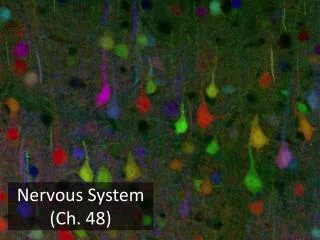
The nervous system consists of three main components: the brain, spinal cord, and nerves. Its primary functions include sensation, integration, and motor response. The central nervous system (CNS) comprises the brain and spinal cord, while the peripheral nervous system (PNS) surrounds them. Neurons are the fundamental units of the nervous system, responsible for carrying messages via nerve impulses. This system features various neuron types, including sensory, motor, and interneurons, which facilitate responses to stimuli. Additionally, neuroglial cells support neuron function and maintain homeostasis.

Understanding the Nervous System: Structure, Function, and Neurons
E N D
Presentation Transcript
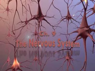
Ch. 9 The Nervous System
The Nervous System It is comprised of 3 basic components Brain Spinal cord Nerves
General Functions of the Nervous System • Sensation • Monitors changes/events occurring in and outside the body. Such changes are known as stimuli and the cells that monitor them are sensoryreceptors. • Integration • The parallel processing and interpretation of sensory information to determine the appropriate response (Conscious or subconscious) • Reaction • Motor output or the activation of effectors such as muscles or glands (typically via the release of neurotransmitters).
Organization of the Nervous System 2 big initial divisions: • Central Nervous System • The brain + the spinal cord • The center of integration and control • Peripheral Nervous System • The nervous system outside of the brain and spinal cord • Consists of: • 31 Spinal nerves • Carry info to and from the spinal cord • 12 Cranial nerves • Carry info to and from the brain
What is the basic functional unit of the nervous system? • Neuron! • What does the neuron do? • Carries messages throughoutthe body • How does it carry the messages? • By conducting electrical signals • What are these signals called? • Nerve impulses!
Neuron Anatomy • Three parts to a NEURON: • Cell body: Large, central portion of the neuron where all organelles are located. • What is its job? • Interpret incoming signals Cell Body
Neuron Anatomy Dendrites 2. Dendrites: Short, highly branched fibers • What is its job? • Carries impulses toward the cell body • Referred to as the afferent process
Neuron Anatomy 3. Axon: Long, slightly branched fiber • What is its job? • Carry impulses away from the cell body • Referred to as the efferent process
Neuron Anatomy Three parts of the Axon: • Axon hillock: site of initiation of an action potential (point where axon and cell body meet) • B. Axon fiber: the main portion of the axon • C. Axon terminal:branched end of the axon (point of communication with other cells)
The Axon Axon Hillock fiber Axon Terminals
Structure of a Typical Neuron Dendrite Axon terminal Cell body Nodes of Ranvier Axon Myelin sheath Nucleus Neuron Anatomy Additional Parts of a Neuron
1 6 2 3 5 4 7 Neuron anatomy What do you remember? dendrites Myelinsheath nucleus Direction of impulse Cell body axon Nodes ofRanvier Axonterminals
3 Types of Neurons A. Sensory Neurons: Receive incoming stimuli • Five types of sensory neurons: • Thermo-receptors • Mechano-receptors • Chemo-receptors • Photo-receptors • Pain-receptors
Thermo-receptors • Location: • Skin • Hypothalamus • Body Core • Function: • Sensation of hot and cold • Detects change in body core temp.
Mechano-receptors • Location: • Skin • Skeletal muscle • Inner ear • Function: • Touch • Pressure • Muscle movement • Motion • Sound
Chemo-receptors • Location: • Nose • Tongue (taste buds) • Blood vessels • Function: • Smell • Taste • Detects levels of CO2 in blood
Photo-receptors • Location: • Eyes • Function: • Allow vision thru detection of light
Pain-receptors • Location: • Everywhere, except the brain • Function: • Sensation of pain • Detects chemicals released by damaged cells
Three Types of Neurons B. Motor Neurons: Carry impulses to muscles and glands Cause a response to some stimuli C. Interneurons: Connect sensory and motor neurons Allow for quick response (reflex action)
Structural diversity in neurons • Multipolar- many dendrites, one axon • Most neurons in CNS • Bipolar- one dendrite, one axon • Sensory organs • Unipolar- sensory • Axon termini extend into CNS
White matter Gray matter Neurons • What is grey matter? • Collective cell bodies and dendrites of all neurons • What is white matter? • Myelinated nerve fibers • Axons of all neurons • Can be approximatelyone meter in length
What is a Nerve? • Bundle of axonsheld together by connective tissue. • What color isa nerve? • White! • Why? • Because axons are white matter and they composenerves
How are nerves held together? • Connective tissue • What is this connective tissue called? • Neuroglial cells (nerve glue) • Approximately half of the volume of the brain is composed of neuroglial cells • Most brain tumors develop in mesoglial cells – NOT neurons
What do neuroglial cells do? • Support the axons • Insulate the electrical impulses • Like electrical tape insulates electric wires this prevents “leaking”of electric signals
Four types of neuroglia in CNS • Oligodendrocytes • Myelinating cells • Astrocytes • Connects neurons and blood vessels together • Microglia • Phagocytes (from bone marrow) • Ependymal cells • Line ventricles of brain; produce cerebrospinal fluid (CSF)
Neuroglia of the PNS • Schwann cells • Myelinating cells • Help direct axon regeneration • Satellite cells • Support, protection, regulation of molecular exchange • “Filter out” other stimuli
The Nerve Impulse • Resting potential – the charge that exists across a neuron’s membrane while at rest. • -70 mV. • This is the starting point for an action potential.
The Nerve Impulse • A nerve signal or action potential is an electrochemical message of neurons. • An all-or-none phenomenon – either the fiber is conducting an action potential or it is not. • Across its plasma membrane, every cell has a voltage called a membrane potential. • The inside of a cell is negative relative to the outside.
The Nerve Impulse • Neuron at rest – active transport channels in the neuron’s plasma membrane pump: • Sodium ions (Na+) out of the cell. • Potassium ions (K+) into the cell. • More sodium is moved out; less potassium is moved in. • Result is a negative charge inside the cell. • Cell membrane is now polarized.
Sodium-Potassium Exchange Pump • Na+ flows into the cell during an action potential, it must be pumped out using sodium pumps so that the action potential will continue.
The Nerve Impulse • A nerve impulsestarts when pressure or other sensory inputs disturb a neuron’s plasma membrane, causing sodium channels on a dendrite to open. • Sodium ions flood into the neuron and the membrane is depolarized – more positive inside than outside.
The Nerve Impulse • This moving local reversal of voltage is called an action potential. • A very rapid and brief depolarization of the cell membrane. • Membrane potential changes from -70 mV to +35 mV. • After the action potential has passed, the voltage gated channels snap closed and the resting potential is restored. • The membrane potential quickly returns to -70 mV during the repolarization phase. • An action potential is a brief all-or-none depolarization of a neuron’s plasma membrane. • Carries information along axons. • An action potential is self-propagating – once started it continues to the end.
Synapses: Junctions Between Nerves • Eventually, the impulse reaches the end of the axon. • Neurons do not make direct contact with each other. • The junction between the axon of one neuron and the dendrite of the next is called a synapse .
Synaptic Pathways • Presynaptic neurons bring action potentials toward the synapse. • Postsynaptic neurons carry action potentials away from the synapse. • A synaptic cleft is the small gap between the two neurons.
Neurotransmitters • Chemical messengers called neurotransmitters carry the message of the nerve impulse across the synapse.
Neurotransmitters • Neurotransmitters are released into the synapse and bind with receptors on the postsynaptic cell membrane, which cause ion channels to open in the new cell.
Reflex Arc • A simple reflex produces a very fast motor response to a stimulus because the sensory neuron bringing information about the stimulus passes the information directly to the motor neuron.
Reflex Arc • Usually, there are interneurons between sensory and motor neurons. • An interneuron may connect two neurons on the same side of the spinal cord, or on opposite sides.
The Central Nervous System • Meninges – are membranes that protect the brain and spinal cord • Dura mater (outermost layer) • Arachnoid membrane ( middle layer) • Pia mater (innermost layer)
The Central Nervous System • Cerebrospinal Fluid (CSF) • Located between the arachnoid mater and pia mater is an area called the subarachnoid space • Continuously secreted from specialized cells (ependymal cells) in the choroid plexus in ventricles • Functions: Physical and chemical protection of the CNS
Spinal Cord • Slender structure that is continuous with the brain • Descends into the vertebral canal and ends around the level of the first or second lumbar vertebra. • Function of the spinal cord is to carry sensory information to and from the brain • 31 spinal segments: • 8 cervical segments • 12 thoracic segments • 5 lumbar segments • 5 sacral segments • 1 coccygeal segment
Spinal Cord - Ascending and Descending Tracts • Ascending tracts - carry sensory information up to the brain • Descending tracts - carry motor information down from the brain to muscles and glands
The Brain Four Parts: Cerebrum Diencephalons Brain stem Cerebellum
The Brain - Cerebrum • Largest part of the brain • Two halves cerebral hemispheres • Thick bundle of nerve fibers called the corpus callosum connect the two hemispheres • Lobes • Frontal • Parietal • Temporal • Occipital • Cortex • Ventricles
The Brain - Diencephalons Located between the cerebral hemispheres and is superior to the brain stem • Thalamus - relay station for sensory information that heads to the cerebral cortex for interpretation • Hypothalamus - maintains balance by regulating many vital activities such as heart rate, blood pressure, and breathing rate.
The Brain - Brain stem • Midbrain - controls both visual and auditory reflexes • Pons - regulates breathing Connects the cerebrum to the spinal cord • Medulla oblongata - controls many vital activities such as heart rate, blood pressure, and breathing