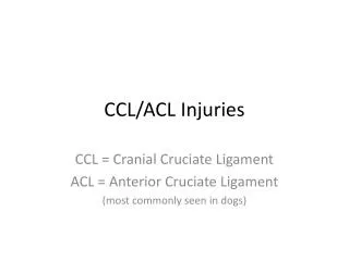
CCL/ACL Injuries
CCL/ACL Injuries. CCL = Cranial Cruciate Ligament ACL = Anterior Cruciate Ligament (most commonly seen in dogs). What is the ACL/CCL?. It’s one of the two ligaments connecting the femur and tibia bones. These ligaments are there to keep the bones from rubbing across each other.

CCL/ACL Injuries
E N D
Presentation Transcript
CCL/ACL Injuries CCL = Cranial Cruciate Ligament ACL = Anterior Cruciate Ligament (most commonly seen in dogs)
What is the ACL/CCL? • It’s one of the two ligaments connecting the femur and tibia bones. • These ligaments are there to keep the bones from rubbing across each other.
How does the injury occur? • Specifically – the knee twists too much and the ligament ruptures. • Examples: • Slipping on a floor • Excessive running • Trauma (i.e. hit by car) • Degeneration of the ligament • Obesity
Breeds Affected • These breeds are seen to have increased risk of degeneration. • Labrador Retriever • Newfoundland • Rottweiler • BichonFrise • St. Bernard • Boxer • West Highland White Terrior • Remember: ANY dog can rupture their ACL/CCL • Most that are predisposed will get it in both knees. • In small breeds, a luxating patella may predispose them.
Symptoms of these injuries • Variable lameness on one or both hind feet • Lameness in affected limb (especially after exercise) that gets better with rest • Abnormal posture • Reluctance to run, jump, or rise from sleep. • Morning stiffness • When sitting, one leg sticks out to one side • Swelling around the knee joint • Slight deterioration of the muscles on the affected limb *If symptoms are very minor, it may be because of slight deterioration.
How is it diagnosed? By watching and touching the animal. With Radiography Only used to access the amount of arthritis present in the knee joint. Picture taken from University of Liverpool website • Veterinarian views a limp with paw slightly touching the ground • Dog will not put any weight on the affected foot. • Veterinarian manipulates the affected knee feeling for ‘drawer movement’ • The movement of the femur across the tibia • This is the only way to confirm the presence of rupture. • http://www.youtube.com/watch?v=9jg9E2nBt_E
How X-rays are used for this injury X-ray technique: Area that x-ray is taken at to get view of ACL/CCL = the stifle/knee joint Best view is lateral but CaCr also necessary. Measurement taken at the area of the widest part of stifle joint. Beam centered over the stifle joint. Veterinarian will measure the slope of the tibia to help choose which surgery to do • Important because they will help evaluate the secondary conditions • Osteoarthritis • Joint cartilage injury • Accumulation of fluid around the joint. Picture taken from University of Liverpool website.
How to fix this injury… • Surgery is the best most definitive option for medium to large size dogs. • There are various types of surgery. • Surgery will correct the ligament problem only • Majority of the time will also have to treat the accompanying issues • Arthritis • Joint fluid • But these problems may not happen for years. • Nylon bands can be used to correct a small dog’s rupture. • Some claim restricted activity for a long period of time after diagnosis will allow the joint to realign itself.
Surgical Correction Options • 1. TPLO – Tibial Plateau Leveling Osteotomy • Most recommended for large breed dogs. • Procedure: • Surgeon checks the cartilage of the knee to determine if it is also torn • There is a cut made into the plateau of the tibia • It is rotated to make the slope more level with the femur • A plate and screws are inserted to make sure that it stays in place. • Much of the success of this option depends on the owners post-op care.
Surgical Correction Options • 2. TTA – Tibial Tuberosity Advancement • a slightly less invasive procedure with slightly quicker recovery time • Results very similar to the TPLO option • Procedure: • Cut is made into the front part of the tibia (the tuberosity) • This part is pushed forward to remove the abnormal sliding of the bone • A specialized bone spacer in placed in the space that was created. Plate and screws also used to secure the bone in place.
Nylon Band Treatment • Most commonly used in small dogs and cats • A suture material made of nylon is passed between the back of the femur to the tibia crest • Scar tissue develops over time to stabilize the joint
Prognosis? • With proper care, animal will return to regular activity. • Will most likely need to be on NSAIDs for the osterarthritis. • If only one side was affected/corrected the other one is more likely to eventually rupture
Sources: • Degner, Daniel DACVS. “Cranial Cruciate Ligament Rupture – Lateral Fabellar Technique (Extrascapular Technique).” Vet Surgery Central Inc. 8 September 2010. http://www.vetsurgerycentral.com/cruciatelrt.htm. • Degner, Daniel DACVS. “Tibial Plateau Leveling Osteotomy.” Vet Surgery Central Inc. 8 September 2010. http://www.vetsurgerycentral.com/tplo.htm. • Degner, Daniel DACVS. “TTA for Cranial Cruciate Ligament Rupture in Dogs.” Vet Surgery Central Inc. 8 September 2010. http://www.vetsurgerycentral.com/ortho_TTA.htm. • Innes, John RCVS. “Cruciate Ligament Rupture.” University of Liverpool Small Animal Teaching Hospital. 8 September 2010. http://www.liv.ac.uk/sath/conditions/cruciate.htm. • Nash, Holly DVM, MS. “Ruptured Anterior Cruciate Ligament.” Pet Education.com. Doctors Foster and Smith. 8 September 2010. http://www.peteducation.com/article.cfm?c=2+2084&aid=474.