Download

1 / 92
1.11k likes | 2.41k Vues
Spinal Cord Injury. Neurosurgeon Yoon Seung-Hwan. General Principles of Spine Injuries. Epidemiology. the fourth leading cause of death in the US C-spine injuries MVAs - 50% falls - 25% sports activities - 10% 60% of all spine injuries in children;

E N D
Spinal Cord Injury Neurosurgeon Yoon Seung-Hwan
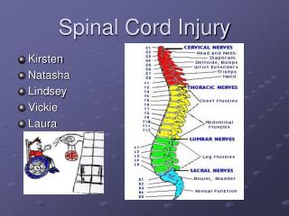
Epidemiology • the fourth leading cause of death in the US • C-spine injuries MVAs - 50% falls - 25% sports activities - 10% • 60% of all spine injuries in children; upper cervical spine. • C5-C6 is the most commonly injured level in adults.
Stability • Definition 1. The ability of the spine under physiologic loads to prevent displacements which would injure or irritate neural tissue. 2. Instability is the loss of the ability of the spine to tolerate physiological loading without incurring neurological deficit, pain, or progressive structural deficit.
Initial evaluation and treatment • ABC's • Stablization for transport don't move patient before stabilization of cervical spine. • In hospital • Usual trauma protocol • Traction rule of thumb is 5 pounds per spinal level above the fracture/dislocation. 60-80 pounds are usually the upper limit in any case.
Pharmacologic treatment of SCI 1.Hypertension adequate volume and normotension 2. Methylprednislone within 8 hours of the SCI protocol 30mg/kg initial IV bolus over 15 minutes followed by a 45 minute pause, and then a 5.4 mg/kg/hr continuous infusion. 3. Naloxone 4. Triliazad
Plain Films • AP, lateral, obliques • open mouth • Swimmer's view
Upper limits of prevertebral soft tissue level of C1: 10 mm level of C4: 7 mm level of C6-7: 20 mm
Inter-spinous process widening (on AP radiographs) More than 1.5 times the interspinous distance at the levels above and below is abnormal.
C1-C2 distance between posterior cortex of C1 arch and odontoid is maximally 3 mm in adults and 4.5 mm in children. • Normal spinal canal diameter is 17 ± 5 mm Stenosis is present if <13 mm.
anterior subluxation of 3 mm of one body on another (or >20% of the AP distance) indicates instability. • angulation greater than 11° is suggestive of instability.
Other Investigations • flexion and extension views, tomograms CT, CT/myleogram, and MRI • MRI essential for suspected spinal cord injury all soft tissues (disk and ligamentous structures) are shown in much better detail.
Odontoid Anatomy • Steele's rule of thirds The dens, subarachnoid space, and spinal cord each occupy 1/3 of the area of the canal at the level of the atlas.
Ligaments External 1. posterior occipito-atlantal ligament (ligamentum flavum ends at C1). 2. anterior occipito-atlantal ligament. 3. ligamentum nuchae. 4. anterior longitudinal ligament.
Internal 1. cruciform ligament( transverse ligament ) 2. accessory ligaments 3. apical ligament 4. alar ligaments 5. posterior longitudinal ligament ( tectorial membane )
Types of injuries • Atlanto-occipital dislocation. • Condylar fractures. • Atlanto-axial dislocation. • Atlas fractures. • Odontoid fractures. • Hangman's fractures. • C3 fractures.
Atlas fractures • cervicomedullary junction remains unchanged because of capacity of spinal canal. • Jefferson’s fracture
Clinical Features • isolated C1 fractures rarely have associated cord injury. • symptoms neck tenderness need neck support pharyngeal protuberance. dysphagia.
Imaging • Spence's Rule If, on plain films, the distance of excursion of lateral masses is 7 mm or more, there is a transverse ligament rupture.
Treatment • Halo immobilization in virtually all cases except when Spence's rule exceeded
Odontoid Fractures • Odontoid fractures are the M/C fractures of C2.
Epidemiology • about 10-15% of all cervical spine fractures. • In children, these consitute about 75% of all C-spine injuries.
Clinical Features • need high index of suspicion in all trauma patients • many signs and symptoms are non-specific • vertebral artery compression may cause brain stem ischemic symptoms. • most patients unwilling to go from supine to sitting position without supporting their heads with their hands.
Imaging • open mouth views • tomography or CT • saggital and coronal reconstructions have superseded tomography
Treatment and results • Halo traction, maximum of 5-10 pounds to achieve reduction • Surgery Type I : no fusion required Type III : no fusion required (>90% fuse with Halo immobilization) Type II : several factors important in decision making
1. Patient age. sixty years of age is the usually quoted cutoff for conservative management. 2. Displacement If >6 mm and >60 years, 85% nonunion rate. 3. Age of fracture greater than 2 weeks seems to be the cutoff for high union rate
Techniques for C1/C2 Fusion • C1/C2 fusions can be considered even if there is a unilateral fracture of the atlas ring, or unilateral fracture of the lateral mass of the atlas.
Hangman's Fracture • When submental knots with a measured corporal drop are used to hang someone, a classic hangman's C2 fracture is the result. • Pathophysiology and Classification Hangman's fracture involves a bilateral arch fracture of C2 (pars interarticularis i.e, the pedicle) with variable C2 on C3 displacement.
Clinical Features • not highly specific symptoms, diffuse neck pain with stiffness • relative sparing of the spinal cord because of the capacious bony canal.
Treatment • type I may be treated in a rigid collar for 12 weeks. • remainder are all treated initially with Halo immobilization. • up to 5% will eventually require surgery.
Surgical Indications • inability to reduce fracture • failure to maintain reduction in Halo vest. • C2-3 disk herniation with spinal cord compromise. • established non-union (late)
Surgical Procedures 1. C1-C3 arthrodesis 2. C2 pedicle screws. 3. C2-C3 anterior discectomy with fusion and plate.
Classification I. flexion-dislocationII. flexion-compressionIII. compression burst IV. extension
Criteria for Instability • White and Panjabi also concluded that horizontal motion between vertebrae should not exceed 2.7 mm (3.5 mm with standard X-ray magnification), and angular motion should not exceed 11 mm.
Three column model • failure of 2 of the 3 columns implies that there will be instability. • 1. anterior column 2. middle column 3. posterior column
Flexion Dislocation Injuries A) unilateral facet subluxation B) bilateral facet dislocation
Indications for surgery 1. failure to obtain reduction 2. failure to maintain reduction 3. pseudoarthrosis 4. purely ligamentous injury 5. re-subluxation 6. any patient with jumped facets