Spinal cord injury
120 likes | 446 Vues
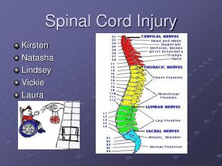
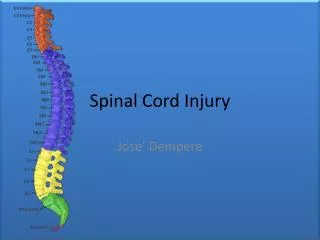
Spinal cord injury. Jessica Ryu , T4 Tulane University School of Medicine. Anatomy. Anterior spinal artery, 2 posterior spinal arteries All 3 receive contributions from the radicular branches 4-10 radicular branches which arise from the vertebral, cervical, intercostal, lumbar arteries

Spinal cord injury
E N D
Presentation Transcript
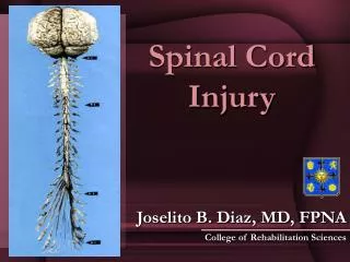
Spinal cord injury Jessica Ryu, T4 Tulane University School of Medicine
Anatomy • Anterior spinal artery, 2 posterior spinal arteries • All 3 receive contributions from the radicular branches • 4-10 radicular branches which arise from the vertebral, cervical, intercostal, lumbar arteries • Anterior spinal supplies 2/3 of spinal cord (motor function) • Posterior arteries supply posterior columns and horns
Anatomy Continued • Cervical and superior thoracic region: derived from cervical branches of the vertebral and ascending and deep cervical arteries • Middle and lower thoracic cord: radicular arteries less prominent • Lower thoracic and lumbar cord: T7-conus blood supply is artery of Adamkiewicz • Greatest susceptibility to cord ischemia: thoracic region
Cervical Spine Injuries • Spinal shock, immediate, lasts for hours to about a month • Flaccid paralysis • Bradycardia, hypotension and EKG changes • Alveolar hypoventilation, hypoxemia and decreased ability to protect airway • Management: • Induction: awake or IV rapid sequence (awake intubation is safest) • Awake: nose is cocainized, oropharynx sprayed with 4% lidocaine, superior laryngeal nerve blocked by injection, recurrent laryngeal can be blocked by injection but in full stomach situation that is probably not advised (if RLN not blocked, cough ability retained)
Cervical Spine Injuries Continued • Important levels: • Diaphragm • Patients don’t survive with injuries above C2 • Important note: patients should be positioned for surgery before they are put to sleep if they have an unstable C-spine
Paralysis • Two stages: flaccid and spastic • Flaccid: 1-4 weeks, manifested by total absence of neuro function below lesion, usually characterized by spinal shock • Spastic: occurs after 4 weeks, manigested by motor hyperreflexia and autonomic hyperreflexia • Problems experienced by paraplegics: bowel, bladder, anemia, dehydration • Spinal anesthesia is a good choice in paraplegics (blocks afferent impulses) • To evaluate level of anesthesia in paraplegic test for sympathogalvanic response
Monitoring • Motor injury detection • Evoked potentials: somato-sensory evoked potentials provide ability to monitor sensory pathway functional integrity • Wake up Test
Important Facts to Remember • Flaccid paralysis (hypovolemia, bradycardia, increased sensitivity to anesthetics) • Ventilation problems and increased risk of gastric aspiration • Hyperkalemia (muscle membrane becomes chemically active – 1 day to 1 year) • Hypothermia (no temp regulation below level of lesion) • Renal insufficiency (risk of infection)
Important Facts to Remember • Unstable thoracic or lumbar spine injuries: patients can be put to sleep on their beds and then moved • Sux contraindicated for about 1 day- 1 year after injury (causes release of K+ from motor end plate membrane and the muscle membrane after spinal cord injury is abnormal)