Spinal Cord Injury
390 likes | 1.26k Vues
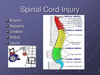
Spinal Cord Injury. Sarah Crosby June 2010. C-Spine. Epidemiology. C-spine injuries occur in 2.0-6.6% of blunt trauma patients Co-existing head injury increases the incidence of C-spine injury to 10%

Spinal Cord Injury
E N D
Presentation Transcript
Spinal Cord Injury Sarah Crosby June 2010
Epidemiology • C-spine injuries occur in 2.0-6.6% of blunt trauma patients • Co-existing head injury increases the incidence of C-spine injury to 10% • Injury to the cervical spinal cord in the absence of fracture occurs in 0.07-0.7% of trauma admissions • Missed or delayed diagnosis of cervical spine injury occurs in 4-8% • Results in 10 x the incidence of secondary neurologic deficit compared to early diagnosis • Of the patients with missed or delayed diagnosis most have decreased GCS, hypotension or critical injuries • Obtunded patients unable to assist in demonstrating neurology • Lifetime healthcare and living costs for a quadriplegic patient $2.4-3.1 million AUD
EAST Guidelines (Eastern Association for the Surgery of Trauma) • 1998 guidelines for evaluation of c-spine injury in trauma patients • 2009 update • CT largely replacing plain x-rays • Clinical clearance remains standard in awake, alert patients with no neurologic deficit, distracting injury, neck pain or tenderness • Cervical collars should be removed ASAP • Controversy still exists regarding cervical spine clearance in the obtunded patient • Who needs imaging • What imaging • How to exclude significant ligamentous injury
Removal of Cervical Collars • As soon as feasible (Level III) • Early removal is associated with: • Decreased collar related pressure ulcers • Skin breakdown 6.8% after 24h • Decreased ICP • Fewer ventilator days • Fewer ICU and hospital days • Decreased incidence of delerium and pneumonia
Penetrating Brain Injury • Immobilisation is not indicated unless the trajectory suggests direct injury to the cervical spine
Clinical Clearance of the C-spine in the awake patient • In awake, alert trauma patients without neurologic deficit or distracting injury, who have no neck pain or tenderness with full range of motion of the cervical spine, imaging is not necessary and the collar can be removed (Level II) • Meta-analysis of clinical clearance of the asymptomatic C-spine (2010) • GCS 14-15 • No posterior cervical tenderness • No neurological deficit • No dangerous mechanism • No distracting Injury • Normal range of motion without pain or neurology Sensitivity 98.1% NPV 99.8%
NEXUS Study Validated 5 Criteria for Low Probability of C-Spine Injury (No imaging required if meet all criteria) • No posterior midline C-spine tenderness • No evidence of intoxication • Normal level of alertness • No focal neurologic deficit • No painful distracting injury Long bone fracture, visceral injury requiring surgical consultation, large laceration, degloving injury, crush injury, large burns Hoffman et al. Validity of a set of clinical criteria to rule out injury to the cervical spine in patients with blunt trauma. NEJM 2000;343:94-99.
Canadian Cervical Spine Rule Study 1. Are there any high risk factors present that require imaging GCS 15 YES NO 2. Are there any low risk factors present to allow for safe assessment by using active range of motion NO Image C-Spine NO YES 3. Can the patient actively rotate the neck 45º left and right No imaging of C-spine needed YES Stiell, I. et al. The Canadian C-spine rule for radiography in alert and stable trauma patients. JAMA 2001;286:1841-1848
High risk factors that require imaging Age ≥65 yo Dangerous mechanism of injury Fall from 1m (5 stairs) Axial load to the head (eg. Diving) MVA- high speed (>100kph, rollover, ejection) Motorised recreational vehicles Bicycle collision Paraesthesia in extremities 2. Low risk factors that allow safe assesment of range of motion Sitting position in the emergency department or Simple rear end MVA or Ambulatory at any one time or Delayed onset of neck pain or Absence of midline C-spine tenderness Canadian Cervical Spine Rule Study
NEXUS Sensitivity 90.7% Specificity 36.8% Radiography Rate 55.9% CCR Sensitivity 99.4% Specificity 45.1% Radiography Rate 66.6% Clinical Clearance of the C-Spine in the Awake Patient Stiell IG, et al. The Canadian c-spine rule versus the nexus low-risk criteria in patients with trauma. NEJM 2003;349:2510-2518
Radiographic Examination of the C-Spine • Plain C-spine X-Rays • 3-view (lateral, AP, odontoid) • Supplemented by swimmers views and CT for poorly visualised areas • CT C-Spine • Occiput to T1 with saggital and coronal reconstruction • More time efficient and cost-effective in moderate and high risk cases • More accurate • Sensitivity 98% (vs c-spine x-ray 52%)
Radiographic Examination of the C-Spine • Indicated for: • Pain or tenderness in the neck • Neurological deficit • Altered mental status • Distracting Injury • The Primary screening modality is axial CT from the occiput to T1 with sagittal and coronal reconstructions (Level II) • Plain radiographs contribute no additional information and should not be obtained (Level II) • If there is neurological deficit attributable to a c-spine injury an MRI should be obtained
Neck Pain with negative CT in the neurologically intact patient 3 options (Level III) • Continue collar • Remove collar after negative MRI (<72h) • Remove collar after negative and adequate flexion extension films • Picks up c-spine instability in 6.75-8% of normal c-spine films • Incidence of isolated ligamentous injury is rare (0.6% of traumatic c-spine injuries)
C-spine Clearance in the Obtunded Trauma Patient with a Negative CT C-Spine and no Gross Neurological Deficit EAST Recommendations 2009 • Flexion/Extension radiographs should NOT be performed (Level II) • The risk/benefit ratio of obtaining an MRI in addition to CT is not clear (individualise to each institution) (Level III) • Options are: • Continue cervical collar immobilisation until a clinical examination can be performed • Remove the cervical collar on the basis of CT alone • Obtain an MRI and if negative the collar can be safely removed (Level II)
? MRI in addition to CT? • Incidence of ligamentous injury with negative CT c-spine is very low (<5%) • Incidence of clinically significant injury is even lower (<1%) • Expensive • Difficult in the intubated patient • Limited availability • More sensitive for identification of soft tissue injuries (Gold standard for spinal cord injury) • Not reliable for identifying bony injury
SCIWORA Significant Cord Injury without obvious radiological abnormality • Incidence 3-5% (x-ray/CT) • Higher incidence in paediatric population (34.8%) • The relatively large size of the head • inherent skeletal mobility • cord vulnerable to damage • Higher incidence above 60 yo • Posterior vertebral spurs due to spondylosis • Ligamentum flavum bulging due to loss of disc height • Risk of central cord syndrome after hyperextension injury
Thoracolumbar Spine Trauma • 4.4% of trauma patients have TLS fracture • 19-50% of these fractures are associated with spinal cord damage • Higher incidence of neurologic deficit when fracture identification was delayed (10.5% vs 1.4%)
EAST Recommendations (2007) • Level II Guidelines • Trauma patients should be examined by a qualified attending physician • Trauma surgeons, emergency physicians or spine surgeons (neurosurgery or orthopaedics) • Trauma patients who are awake, without any evidence of intoxication, with normal mental status, neurologic and physical examinations may be cleared clinically
EAST Recommendations (2007) • Radiographic Examination is required for: • High energy mechanism of injury • Falls from >10 feet • MVA/MBA crash with or without ejection • Pedestrians struck • Assault, Sport or Crush injury • Bicycle injury • Concomitant cervical spine fracture • Altered mental status, intoxication • Distracting injuries • Neurologic deficits • Spine pain or tenderness on palpation
Mode of Imaging of TLS • Multidetector CT with axial reconstruction is superior to plain films for screening of TLS for bony injury (II) • CT scout films can be used for spine assessment (II) • CT scan may be associated with less overall radiation exposure than plain films (III) • Plain films are adequate for the examination of the TLS if the patient does not require CT scan for any other reason (III) (Not if they have a major trauma mechanism) • MRI is indicated for patients with neurologic deficits, abnormal CT scans or clinical suspicion despite normal radiographic evaluation suggesting an unstable injury (III) • Early decompression of traumatic lesions improves outcome
Plain Film vs CT of TLS • Ballock et al. (1992) • plain radiography of the thoracolumbar spine would have missed 25% of fractures • Gestring et al. (2002)- CT protocol for examining TLS • Anterior, posterior and lateral scout films and axial images • 100% sensitivity and specificity • Hauser et al. (2003)-prospective study 222 patients • Plain radiography of the TL spine vs Helical CT (5mm images) • CT scan accuracy 99% vs plain radiographs 87% • CT could also differentiate acute vs old # • Sheridan et al. (2003) • Reformatted helical T (2.5mm images) vs plain x-ray • Sensitivity for Thoracic #- CT 97% vs x-ray 62% • Sensitivity for Lumbar #- CT 95% vs x-ray 86%
“Clearing” the Spine • The ultimate evaluation of all radiographic studies is the responsibility of the attending radiologist. • Other persons qualified to interpret TLS radiographs • Trauma surgeon • Emergency medicine physician • Neurosurgeons • Orthopaedic spine surgeons • These may clear the spine after interpretation of the images and clinical evaluation of the patient
Obtunded Patient • No level I evidence • Level II • Multidetector CT with axial reconstruction is superior to plain films for screening of the TLS for bony injury • Level III • The obtunded patient (intoxicated or head injury) presenting to a centre without CT scan capability should be transferred to the nearest available trauma centre
Primary Management A - Airway control with C-spine immobilisation B- Avoid hypoxaemia- will further worson the prognosis of an injured spinal cord - Injury above C5 will cause respiratory insufficiency - 50% of C3 tetraplegics need permanent ventilation C- Need to minimise secondary ischaemic injury to the cord - Aim MAP > 100mmHg - SCI above C6 associated with loss of cardiac sympathetic supply leading to hypotension & bradycardia - Loss of sympathetic vasoconstriction leads to vasodilation & venodilation - relative hypovolaemia needing plasma volume expansion and vasoconstrictors
Secondary Survey of Spine • Assessment of the cervical soft tissue for swelling • Log roll and palpation of the spinous processes of the entire spinal column • Neuro exam • Motor, sensory and reflexes • Perianal sensation, rectal sphincter tone and sacral reflexes • Absence of the bulbocavernosus reflex indicates spinal shock • Signs of SCI in unconscious patient • Response to pain above but not below a level • Flaccid areflexia in arms/legs • Elbow flexion with the inability to extend (cervical SCI) • Paradoxical breathing • Inappropriate vasodilation • Unexplained bradycardia, hypotension • Priapism • Loss of anal tone and reflexes
Spinal Cord Injury • Complete SCI • muscle paralysis • somatic and visceral sensory loss below a discrete segment • Spinal shock • Additional features of • Muscle flaccidity • Absence of tendon reflexes • Vaso and venodilation • Loss of bladder function • Paralytic ileus • Due to temporary loss of somatic and autonomic reflex activity below the neurological level of the injury • Lasts 1-3 weeks
Early Surgical Management of Spine Injuries • Cervical Spine Injuries • Acute stabilisation with a halo ring allows further diagnostic evaluation and treatment of other injuries without further neurologic deterioration • Thoracolumbar Spine Fractures • McLain & Benson compared urgent spinal stabilisation (+/- decompression within 24h) vs early treatment (24-72h) • No statistical difference in outcomes • the urgent group had a non-statistical better neurological recovery • Kerwin et al. • Early stabilisation (<3 days) shortens ICU and hospital LOS but increased number of perioperative deaths and pneumonia • Spinal cord compression with neurologic involvement as a result of spinal malalignment should be remedied as soon as possible (esp facet subluxations or dislocation)
Medical Management of Acute Spinal Cord Injury • Primary Injury • Primary cell death that occurs at time of injury • Due to direct mechanical forces causing structural disruption of neuronal, glial and vascular structures • Secondary Injury (days to weeks) (10% of injury) • Variety of chemical pathways including hypoxia, ischaemia, ionic shifts, lipid peroxidation, free radical production, excitotoxicity, eicosanoid production, protease activation, prostaglandin production and apoptosis • Medical strategies for treatment of SCI are directed at minimising the degree of secondary injury
Spinal Cord Perfusion Pressure • Most important medical management of spinal cord injury is to maintain arterial oxygenation and blood pressure support • Class III evidence to suggest optimising spinal cord perfusion improves clinical outcome • Maintain SBP between 85-90mmHg for first week
Steroids • NASCIS II (1992) • Prospective randomised double blind controlled multicentre trial (487 patients) • High dose Methylprednisolone vs Naloxone Vs Placebo given within 24h of SCI • No difference in neurologic outcome between the 3 groups • Post hoc analysis showed patients treated with Methylpred within 8h had statistically significant improvement in motor and sensory scores at 6 months, but only improved motor scores at 1 year • Steroid group had increased pulmonary emboli, wound infections (non-significant), myopathy • NASCIS III • 48h Methylprednisolone was associated with severe pneumonia, severe sepsis.
GM-1 Ganglioside • Compound normally found in cell membranes of CNS tissue in mammals • Antiexcitotoxic activity • Promotes neuritic sprouting • Potentiates the effects of nerve growth factor • Prevents apoptosis • Initial promising results, not reproduced in long term follow-up • Not used in clinical practice
Future Directives • Stem cell transplantation • Electric Field gradients to influence nerve growth after injury • Neuroprotective strategies • Monoclonal antibodies • Minocycline • Rho inhibitor • Macrophage injection
References Ackland, H. et al. Magnetic Resonance Imaging for clearing the cervical spine in unconscious intensive care trauma patients. J. Trauma 2006;60:668-673 Practice Management Guidelines for identification of cervical spine injuries following trauma- update from the Eastern Association for the Surgery of Trauma Practice Managament Guidelines Committee. J.Como, J.Diaz, M Dunham J Diaz et al. Practice Management Guidelines for the Screening of Thoracolumbar Spine Fracture. J Trauma 2007;63:709-718 Harris, M. et al. The initial assessment and management of the Multiple-Trauma patient with an associated spine injury. Spine 2006;31:S9-S15 Hurlbert, R. Strategies of Medical Intervention in the Management of Acute Spinal Cord Injury. Spine 2006;31:S16-S21 Rechtine G. Nonoperative Management and Treatment of Spinal Injuries. Spine 2006;31:S22-S27 Stiell IG, et al. The Canadian c-spine rule versus the nexus low-risk criteria in patients with trauma. NEJM 2003;349:2510-2518 Stiell, I. et al. The Canadian C-spine rule for radiography in alert and stable trauma patients. JAMA 2001;286:1841-1848 Hoffman et al. Validity of a set of clinical criteria to rule out injury to the cervical spine in patients with blunt trauma. NEJM 2000;343:94-99. Richards, P. Cervical Spine Clearance: a review. Injury, Int. J. Care Injured. 2005;36:248-269 Ed Bersten, A. Soni, N. Oh’s Intensive Care Manual, 5th Edition.2003. Ed Wilson W.et al Trauma- Emergency Resuscitation, Perioperative Anaesthesia, Surgical Management. Vol 1. 2007