Download
1 / 47
521 likes | 1.02k Vues
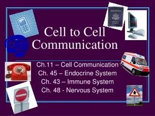
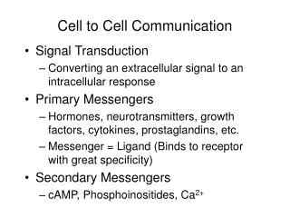
Cell-to-Cell and Cell-to-Matrix Adhesions. Test Your Knowledge: Where would you find the basal lamina ? Proteoglycans? Fibronectin? Name one type of cell-to-cell connection. Name one type of cell to extracellular matrix connection Name one type of cell membrane to cytoskeleton connection.

E N D
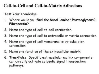
Cell-to-Cell and Cell-to-Matrix Adhesions • Test Your Knowledge: • Where would you find the basal lamina? Proteoglycans? Fibronectin? • Name one type of cell-to-cell connection. • Name one type of cell to extracellular matrix connection • Name one type of cell membrane to cytoskeleton connection. • Name one function of the extracellular matrix • True/False Specific extracellular matrix components can directly activate cytosolic signal transduction pathways.
Cells combine to form tissues. This requires that cells “adhere” to one another to form a functional unit. • Types of adhesion: • Cell-to-cell adhesion • Cell-to-extracellular matrix adhesion;
Types of interactions between adhesion proteins: • Homophilic – adhesion created by interaction between two similar adhesion molecules • Heterophilic – adhesion created by interaction between two different adhesion molecules or between adhesion molecules and cytoskeleton or extracellular matrix proteins • Homotypic – adhesion between similar molecules • Heterotypic – adhesion between different molecules 5 classes of CAMs Not shown: mucins
Cell-to-cell adhesion molecules • 1. Calcium dependent adhesion molecules (cadherins) • Evolutionarily ancient; widely expressed; over 12 different types known • Almost all vertebrate cells express one or more • Structure: Single-pass transmembrane glycoprotein composed of about 700-750 residues • Type of binding: • Types: • Interactive with actin cytoskeleton: Cadherins N; P; R; B; E • Desmosome associated: Desmogleins & Desmocollins • Protocadherins • Location:
E-Cadherin Domains 1 and 2 In Complex With Calcium Two of the 5 tandem repeats of extracelluar region
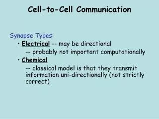
Non calcium dependent adhesion molecules • (NCAMs nerve cell adhesion molecules, ICAMs, and L1) • Evolutionarily ancient; widely expressed • Belong to the immunoglobulin (Ig) superfamily • Structure: single pass, transmembrane proteins which may bind to the cytoskeleton inside cells • Type of adhesion: • Can have both homophilic and heterophilic interactions; • homo – neural specific Ig Cell Adhesion molecules (IgCAMs); • hetero systemic IgCAMs • Functions: • neurite outgrowth, myelination, and • firm adhesion of leukocytes
Selectins • Expressed only in vertebrates; in circulatory cells (endothelium and blood cells) • Transient transmembrane binding proteins (lectins) • In the presence of calcium, bind to specific oligosaccharides on another cell • Structure: single transmembrane polypeptide • Type of adhesion: • Function: extravasation
Cell to extracellular matrix adhesion molecules • Integrins (examples: laminin, fibronectin, fibrinogen) • A family of transmembrane adhesion molecules (usually glycoproteins) that exist in variable activation states • Extracellular matrix receptors on integrins have selective affinity for certain matrix proteins; allows cells to explore their environment • Structure: have an alpha and a beta subunit (heterodimer); alternative splicing has led to 16 different a chains and 8 different b chains • Type of adhesion: • Function: WBC binding to endothelium;
Epithelial tissues – what do they need adhesion molecules for? tight junctions adherens junctions desmosome hemidesmosome
Tight Junctions Function: Protein composition: occludin claudin junction adhesion molecules (JAMs) Cytosolic face
Adherens junctions (adhesion belt); attach to actin Desmosomes; attach to intermediate filaments Focal adhesions Hemidesmosomes
Adherens Junctions Composition: cadherens Function: can contract (with help of myosin) Folding of sheets into tubes during morphogenesis, other folding processes during morphogenesis Binding partners: catenins, and via catenins to cytoskeleton (actin)
desmosome pemphigus
Focal Adhesions Examples: myotendinous junction fibroblast migration in connective tissue
Basal Lamina – extracellular matrix; a sheetlike meshwork underlying or surrounding groups of cells Function :
Components of the basal lamina; produced by cells that rest on it (mainly fibroblasts); sometimes called the basement membrane • Type IV collagen • Laminins • Entactin (nidogen and laminin) • Perlecan
The regulatory factors which impact on matrix synthesis, degradation and function are many and include 'growth factors', cytokines, hormones, vitamins, matrix metalloproteinases (MMPs) and tissue inhibitors of metalloproteinases (TIMPs). Vitamins: C, D Hormones: estrogens, glucocorticoids, Matrix Metalloproteinases MMP: zinc containing enzymes that degrade most molecules of the ECM Tissue Inhibitors of Metalloproteinases TIMP: zinc binding endopeptidases Growth Factors: TGFß promotes cellular movement through matrix, and is involved in imflammation and repair
Proteoglycans – glycoproteins containing covalently linked polysaccharide chains called glycosaminoglycans (GAGs); high viscosity and low compressibility hyaluronan Also: chondroitin or dermatan sulfate, keratan sulfate, heparan sulfate/heparin-
Common structural make-up of GAGs attachment to proteins; proteoglycans or mucopolysaccharides • 95% carbohydrate by weight • Possible functions: • Selective sieve; regulate movement of molecules and cells • Chemical signaling between cells; bind certain growth factors (FGF) to stimulate proliferation in the area; TGFb binds to several core proteins of the proteoglycan group • Bind and regulate proteases and protease inhibitors (may restrict range of action, sterically block activity, provide a reservoir for later release, prolong action, or alter concentration)
GAG Localization Comments Hyaluronate synovial fluid, vitreous humor,ECM of looseconnective tissue large polymers, shock absorbing Chondroitin sulfate cartilage, bone, heart valves most abundant GAG Heparan sulfate basement membranes,components of cell surfaces contains higher acetylated glucosamine than heparin Heparin component of intracellular granules of mast cellslining the arteries of the lungs, liver and skin more sulfated than heparan sulfates Dermatan sulfate skin, blood vessels, heart valves Keratan sulfate cornea, bone, cartilage aggregated with chondroitin sulfates
ECM proteins in connective tissue • Collagen • Proteoglycans • Adhesion proteins • Hyaluronan • Elastic fibers Tendon – dense connective tissue
Type IV collagen • Repeating sequence (Glycine – X –Y)n X = proline Y= hydroxyproline • Left handed helix. N-terminal and C-terminal ends have globular domains (solubility; cleaved when secreted from cell - insoluble) • In ER and Golgi they are glycosylated, OH groups added, S-S links three chains • End result a triple helix
Different types of collagen can co-assemble to form large fibers. Type VI +type I in tendons, form in direction of stress. Type II and Type IX oriented randomly and are in cartilage for strength and compressibility.
Laminins – multiadhesive matrix proteins Function: organization of basement membrane; have binding sites for itegrin receptors (important in embryonic development and tissue remodeling) • Laminins are tightly associated with entactin or nidogen a 150-kD sulfated glycoprotein, which also binds to type IV collagen. As a result of these multiple interactions, laminin, entactin, type IV collagen, and perlacan form crosslinked networks in the basal lamina.
Adhesions between non-epithelial, cells and the extracellular matrix – short and long term adhesions that help in motility • Focal adhesions • Focal contacts • Focal complexes, • 3D adhesions • Fibrillar adhesions • Podosomes
Two conformations of integrins Change in conformation transferred to other proteins scaffolded to internal signaling pathways