Progress in Development of a High-Resolution PET Prostate Imaging Probe
10 likes | 170 Vues
2011 MIC18.M60. N.H. Clinthorne 1 , S. Majewski 2 , A. Stolin 2 , R.R. Raylman 2 , S.S. Huh 1 , A. Studen 3 , M. Grkovski 3 , K. Brzezinski 4 , H. Kagan 5 , D.S. Smith 5 , J. Carr 1 , Z. Chen 1 , E. Salomonsson 1 , A. Yande 1

Progress in Development of a High-Resolution PET Prostate Imaging Probe
E N D
Presentation Transcript
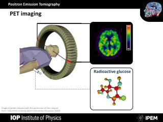
2011 MIC18.M60 N.H. Clinthorne1, S. Majewski2, A. Stolin2, R.R. Raylman2, S.S. Huh1, A. Studen3, M. Grkovski3, K. Brzezinski4, H. Kagan5, D.S. Smith5, J. Carr1, Z. Chen1, E. Salomonsson1, A. Yande1 1Dept. Radiology & Biomedical Engineering, U. Michigan, Ann Arbor, MI USA, 2Advanced Imaging Laboratory, Dept. Radiology, West Virginia University, Morgantown, WV USA 3Jozef Stefan Institute, Ljubljana, Slovenia,4IFIC/CSIC U. Valencia, Valencia, Spain, 5Dept. Physics, 5Ohio State Univ. Columbus, OH USA PET in prostate cancer Double-sided MPPC readout of LYSO for DOI Conventional PET ring Promising as it is, use of PET in clinical management of prostate cancer remains controversial. Small lesions within the prostate are difficult to detect and quantify due to the modest resolution of present scanners and high attenuation of annihilation radiation emanating from the prostate. To address this problem, our collaboration has been investigating construction of endorectal PET magnifying probes to supplement conventional scanner data.* At right is a diagram showing how a high-resolution endorectal detector works in concert with the lower resolution PET ring to provide high spatial resolution in directions parallel to the probe face within the prostate. We have constructed prototype detectors and are presently interfacing them to a partial PET ring for imaging tests. Intensity Profile Excellent resolution close to probe Modest resolution Irradiation As noted previously, good DOI resolution may be desirable as long as it does not significantly reduce the amount of LYSO that can be packed into the probe. Shown above is a prototype DOI detector with an array of 1mm x 1mm x 10mm LYSO optimized for DOI performance. The array is read out by two of the 5 x 5 MPPC arrays with light-spreaders. Intrinsic performance is shown in the accompanying images. Especially relevant is the DOI histogram of front-side vs. back-side signals for a coincidence-collimated 511keV beam moved in 1mm steps. DOI resolution is ~1mm FWHM. Combined performance with PET ring should correspond with red curve in previous plot. Combining front- and back-side signals via geometric mean results in 12.7 keV FWHM energy resolution. Progress in Development of a High-Resolution PET Prostate Imaging Probe High resolution endorectal PET detector (“probe”) DOI Histogram GEOMETRICAL MEAN Intrinsic resolution of probe geometry FWHM 12.7% @ 511keV The intrinsic width of the coincidence line-of-response (LOR) at fractional distance α from the probe detector is given by the following expression where rxrepresents the spatial resolution of each detector in the circumferential and depth directions, θxthe angles of LOR incidence, and D the detector separation in millimeters: Combined Spectrum Next step in probe testing with “full ring” PET For upcoming studies, a complete PET ring will be emulated using the partial ring of BGO block detectors from a CTI 931 PET scanner. Shown at right are the 24 BGO detectors at 500mm radius as well as two high resolution detectors in a magnifying geometry for a small FOV. These inner detectors will be replaced with the LYSO/MPPC probes for upcoming measurements and the FOV size will be increased to ~40cm. Phantoms are rotated to emulate the complete ring. Shown at right is a plot of the intrinsic spatial resolution as a function of distance from a 10mm thick LYSO probe having 1mm circumferential resolution in coincidence with a conventional PET ring with 4mm FWHM detector resolution. Results are shown for a probe having no depth-of-interaction (DOI) resolution and for probes having 1mm and 2mm FWHM depth resolution at several angles of LOR incidence. Note that at near normal incidence (0°) for all probes, resolution improvement can be significant even at 50mm from the probe face. Is depth-of-interaction resolution required in a probe? Maybe. It depends on the angles of incidence of LORs traversing the prostate. In any case, good DOI resolution is desirable. Probe reconstructions showing improved resolution Toward human use Probe measurementgeometry The eventual goal of the project is to test probes in human subjects. To this end, we enlisted students in a Biomedical Engineering capstone design course to determine biocompatible probe sizes, develop appropriate light- and moisture-proof packaging, and evaluate methods of tracking the position and orientation of the probe. Shown at right are several prototype housings. The light colored units were created using 3D printing; the exploded version shows the housing, end-cap, and detector and motion-tracking internals. The dark units are light-proof housings manufactured from black Delrin and can be used with appropriately sized probe detectors. Maximum probe diameter should be < 35mm for “comfortable” use. Rotated 90° CCW from Ring + Probe geometryshown at right Data acquired using the system above provides a demonstration of resolution advantages—and features of limited-angle tomography—in the probe geometry. The drawing above illustrates the imaging geometry of the probe, which had 1.4mm x 1.4mm x 1mm detector elements located ~70mm from the 50mm diameter resolution phantom. Reconstruction from the BGO ring data only results in a low resolution reconstruction. Adding the probe data results in significantly higher resolution “parallel” to the probe face but lower resolution “perpendicular” to the face consistent with the limited-angle nature of the data. Intensity Profiles LYSO / MPPC probes developed at WVU In the past month, two prototype probes have been provided by the WVU Advanced Imaging Laboratory to the lab at Michigan: the non-DOI probe shown directly below and the depth-sensitive probe in the next panel. Probes are constructed using 5x5 arrays of 3mm x 3mm S10362-33-050P Hamamatsu MPPCs (AiT Instruments) shown in the leftmost two images below. For the non-DOI probe (shown disassembled below) a single 5x5 array is coupled via a 2.5mm light spreader and a 24x24 array of 1mm x 1mm x 10mm LYSO crystals. With the exception of the edges, crystal separation is very good as shown at right below. Summary The individual components necessary for demonstrating and evaluating the imaging performance of endorectal prostate probes are quickly coming together. Over the next few months, the LYSO/MPPC instruments provided by the team at WVU will be interfaced to the partial-ring BGO system at Michigan to verify previous predictions from Monte Carlo simulation and analysis. Although we anticipate good performance, much additional development must be done for probes usable in human subjects. Most significant are (1) miniaturizing the probe detector to fit within the 35 mm external diameter constraint, and (2) working with a current manufacturer of PET scanners to create a test instrument. ~25mm 16mm * Funded in part by the US Army Congressionally Directed Medical Research Program under Synergistic Idea Grant W81XWH-09-1-0413