Download

1 / 15
150 likes | 178 Vues
Explore the use of Small Animal PET Systems in biomedical, pharmaceutical, and genetic research. Learn about imaging processes and challenges, with a focus on improving resolution for genetic manipulation studies. See examples of experimental set-ups and results, and discover future work in this innovative field.

E N D
Very High Resolution Small Animal PET Don J. Burdette Department of Physics
What is PET? Answer: Cancerous cells have a higher metabolism than normal cells, thus PET scans detect cancer. Other Uses for PET include detection of Cardiovascular Disease, Alzheimer’s disease, Parkinson’s disease, epilepsy, and other neurological disorders. • PET stand for Positron Emission Tomography • It is a leading medical imaging technique (more than 200,000 PET scans in over 700 institutions performed last year in the US) • Unlike MRI and X-Ray (which image bodily structure) PET creates images of metabolic processes Question: Why is imaging metabolic processes important? Example of a PET image An image of damaged heart (left) compared to a normal heart (right). The damaged heart has undergone a heart attack. Single slice
How does PET work? Photons Electron - + Radioactive Tracer Positron
How does PET work? • Typical Resolution of human full body scans about 10mm • There exist other applications for PET technology outside human imaging
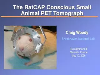
Small Animal PET Systems Uses of Small Animal PET systems: • biomedical, pharmaceutical, and genetic manipulation research • Mice are used as human disease models • A single mouse can be used to tract disease development and treatment (Important for genetically altered specimens) Example of a Small Animal PET image Challenges for Small Animal PET: • Need resolution of less than 1mm • Lower doses, high efficiency Possible Solutions: Transgenic mouse showing cells with albumin gene switched on • smaller detection elements • higher efficiency detectors • Increase solid angle of the detector
MicroPET R4 from University of Michigan’s PET center • Consists of 8 X 8 array of individual LSO scintillation crystals coupled to 64-channel PMT • 15 cm detection ring diameter • Typical System resolution = 1.8 mm • Image of two 1.1-1.2mm capillary tubes filled with Flourine-18 source
Silicon Detectors Characteristics of ideal Silicon detector for use in PET: • Thick detector to increase efficiency (1mm compared to typical 0.3mm thickness) • Small detection pads for excellent spatial resolution • Custom made readout chips including an amplifier, and sample-hold switch • Chips have trigger logic to allow independent silicon operation Silicon Detector composed of an array of 32 X 16 pads 1mm thick by 1.4mm X 1.4mm in area • Small Detection Pixels yield excellent spatial resolution of the absorbed photons • Timing resolution of 200ns (Coincidence Window) Americium-241 spectrum collected by silicon detector
Experimental Set-up In collaboration with the University of Michigan PC Silicon Detector Data Aquisition Module Coincidence Logic Intermediate Board Intermediate Board Collimated Source Si Trigger 1 Si Trigger 2 Si Signal Source Si Signal Mechanical Set-up Si Detector Si Detector
Reconstructing the Data for a Simulated Disk Source Simple Back-Projection of the data (just drawing the lines) Fourier transforming this image into frequency space…
Reconstructing the Data for a Simulated Disk Source • Blue line = unfiltered data • Green line = filtered data • The narrower the peak, the higher the resolution = sharper image Projection of previous image with and without filtering
Reconstructing the Data for a Simulated Point Source Image of Disk Source after filtering and transforming back to position space
Reconstructing the Data for a Simulated Disk Source Original Image Filtered Image
Our small animal PET set-up Resolution of 0.7mm From microPET R4 set-up Resolution of 1.8mm Experimental Results Images of Flourine-18 source contained in two 1.1-1.2mm capillary tubes with a wall thickness of 0.2mm.
Conclusion and Future Work • Our prototype Small Animal PET achieves a system resolution below the 1mm goal demonstrating the usefulness of silicon in Small Animal PET applications. Future Work includes: • Add a stack of silicon detectors to increase efficiency • Improve rate capabilities by decreasing coincidence window This can be accomplished by taking advantage of the Compton scattered photon and using a secondary detector with faster coincidence timing (Silicon timing 200ns coincidence window, timing from some scintillation crystals approach 60ns)
References • Miles N. Wernick and John N. Aarsvold, editors. Emission Tomography: The Fundamentals of PET and SPECT. Elsevier, Acad Press, 2004. • Glenn F. Knoll. Radiation Detection and Measurement. John Wiley and Sons, Inc. 1989. • Christof Knoess. Performance evaluation of the Micropet R4 pet scanner of rodents. Eur J Nucl Med Mol Imaging, 2003. • Special Thanks to Neal Clinthorne, Klaus Honscheid, Harris Kagan, Sang-June Park, and Joseph Regensburger