Reproductive systems in humans
280 likes | 294 Vues
Explore the main parts, functions, and processes of the male and female reproductive systems. Learn about sperm production, ovary functions, and gametogenesis in an easy-to-understand format.

Reproductive systems in humans
E N D
Presentation Transcript
Start by labelling the main parts of the male reproductive organs – use page 100 in your book • Key words Penis, scrotum bladder, pelvis, prostrate gland, urethra, epididymis, testis, seminal vesicle, vas deferens
Annotating the main parts • Testis – secrete the male sex hormones (testesterone) and produce the male gametes (sperm) • Epididymis – a duct which collects and stores spermatazoa from the testis. • Vas deferens – sperm duct – spermatozoa are expelled through this duct. Seminal fluid is added on route. • Exocrine glands – seminal vesicles and prostate glands which secrete seminal fluid (semen) which is nutritive and lubricating along the vas deferens • Semen contains spermatozoa, seminal fluid, mucus and cells lost from the lining • Penis – has erectile tissue which fills with blood to become rigid during sexual intercourse.
Main functions of male reproductive system • To produce and expel motile male gametes (sperm)
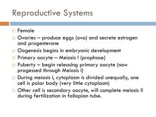
Label the main parts of the female reproductive organs – use page 100 in your book Key words Ovary, cervix, uterus, vagina, rectum, urethra, labia minora, labia majora, clitoris, bladder, rectum, pubic bone, oviduct
Annotating the main parts • Ovaries- site of production of the egg (ova) and secretes the hormones progesterone and oestrogen • Oviducts – move the ova from the ovaries to the uterus (ciliated). Site of fertilization. • Uterus (womb) – site where the fetus implants and develops in the lining known as the endometrium (blood lining) • Vagina – adapted for reception of the penis and passage of the baby during birth.
Main functions of the female reproductive system • Produce gametes (eggs/ ova) • Receive male gametes (sperm) • Provide a suitable place for fertilization and the development of the fetus.
Gametogenesis Production of the gamete cells – sperm and ova
Spermatogenesis • Spermatogenesis – production of the sperm. • This happens in the testis within the seminiferous tubules • Spermatogenesis begins at puberty and normally continues throughout life. • The whole process takes approx 74 days
Outside of seminiferous tubules It starts on the outside of the seminiferous tubules. As the sperm develop they move inwards toward the lumen of the seminiferous tubules. The sperm are eventually pushed into the epididymis and stored until ready for ejaculation into the vas deferens and beyond!! Lumen of Seminiferous tubules
The stages • Primordial germ cells (2N) lining the seminiferous tubules divide by mitosis producing spermatogonium (2N) type 1 • Spermatogonium undergo more mitosis (and some differentiation) to produce primary spermatocytes.(2N) • primary spermatocytes undergo meiosis to produce secondary spermatocytes (n) • secondary spermatocytes undergo meiosis 2 to produce spermatids (n) • Finally the spermatids develop into spermatozoa by a process called spermiogenesis
The sperm cell is 55 to 65 micrometres long • The head contains a nucleus and is covered in an acrosome containing lytic enzymes (to digest the outer coat of the egg) • The midpiece contains many mitochondria needed for energy to help the sperm swim.
Sertoli cells • These cells act as ‘nurse cells’ for the process of spermatogenesis. They provide mechanical and metabolic (energy) support the spermatozoa • They are also phagocytic and ‘eat up’ any excess cytoplasm lost during the process.
Testosterone • Testis also produce testosterone – the male sex hormone. • It is secreted by the leydig cells in the testis • Testesterone controls spermatogenesis as well as many male characteristics such as growth of muscle, voice deepening, behaviour (aggression)
How the brain controls Testosterone • 2 gonadotrophic hromones: Follicle stimulation hormone (FSH) and Interstitial cell stimulating hormone (ICSH) are released by the anterior pituitary gland. • ICSH stimulates the leydig cells to secrete testosterone. • FSH works with testosterone to stimulate spermatogenesis • But testosterone inhibits ICSH which controls the rate of sperm production. • So you can’t make too many sperm!!!
Quick review Q1. Put the following in order • Secondary spermatocyte • Spermatozoa • Primordial germ cell • Spermatogonium • Spermatids • Primary spermatocyte
Answer • 3, 4, 6, 1, 5, 2 • Q2. What process happens between secondary spermatocytes and becoming spermatids? • Meiosis 2 • Q3 what is the process called where spermatids develop into spermatozoa? • Spermiogenesis • Q4. What are the ducts called where the spermatozoa are stored? • Epididymis • Q5 what are the tubes called where spermatogenesis occurs? • Seminiferous tubules • Q6/ what is the name of the nurse cells which support the spermatozoa?
Page 103- Oogenesis – 1.During early fetal development - starts with primordal germ cells then goes through mitosis to form oogonia. • 4/5 months later, some oogonia enlarge and become primary oocytes. Most oogonia degenerate at this time. • 7th month (still before birth) primary oocytes become primordial follicles and begin meisois but then stops until puberty!!!
4. At puberty, once a month, a primary oocyte starts meiosis again. (most primary oocytes degenerate) 5. The follicle containing the primary oocyte enlarges and meiosis 1 is completed. 6. It is now a secondary oocyte. 7. At ovulation the mature follicle (containing the secondary oocyte) ruptures and the secondary oocyte is released.
8. The secondary oocyte (now known as the ovum) moves down the oviduct. 9. If it is fertilized by a sperm then after the head of the sperm has entered the secondary oocyte undergoes meiosis 2 to become the mature ovum. 10. Fertilization can occur. 11. These last 3 stages happen very quickly. Note – all polar bodies degenerate.
The corpus luteum • After ovoulation the ruptured follicle becomes a termpory endocrine structure, the corpus luteum. • The corpus luteum secretes progesterone and oestrogen for up to 12 – 14 days (it gradually gets smaller and smaller. • If fertilization does not occur, it eventually degenerates into a corpus albicans.