Download

1 / 48
480 likes | 691 Vues
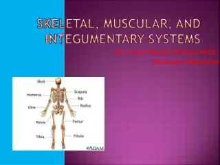
The I ntegumentary System. 1 organ + accessory organs. Skin is an organ Accessory organs Hair follicles Sebaceous glands Sweat glands nails. Like other organs. Skin is made of many tissues Has many functions. Cutaneous Membrane. Another name for skin

E N D
1 organ + accessory organs • Skin is an organ • Accessory organs • Hair follicles • Sebaceous glands • Sweat glands • nails
Like other organs • Skin is made of many tissues • Has many functions
Cutaneous Membrane • Another name for skin • What are the other two epithelial membranes? • Hint: end of chapter 5 Serous Mucous
Functions • Protection • Excretion • Maintain homeostasis • Sensory reception • Blood reservoir • immunity Memorize this!!
2 Distinct Regions • Epidermis • Dermis
Functions • Physical Barrier • Water loss • Injury • Chemicals & microbes
Chemical Barrier • pH of 5-6 • Prevents microorganism growth
Biological Barrier • Langerhan’s cells • Macrophages and mast cells
Excretion • Urea – waste product from metabolizing protein (colorless, odorless). Excreted in sweat and urine. Used in cosmetic products. • Uric acid (guano) – different chemical compound. Leads to kidney stones. Only excreted through urine. Minimal compared to kidneys 99% water
Regulate Body Temperature • Too hot? • Too cold? Vessels constrict and sweating stops RADIATION and DILATION
Nerve Fibers • Associated with muscles, glands and sensory receptors.
Structure of Epidermis BOYS SAY GIRLS LOVE CHOCOLATE
Keritanization • Process that converts basale skin cells into dead, flattened, hardened bags of keratin protein. • http://www.youtube.com/watch?v=OKosGSm7Ps4
Epidermis • Main Job: Protection • Waterproofing • Protect lower layers from trauma • Barrier against microorganisms and chemicals
Dermis • Papillary layer (20%) • Loose areolar CT • Reticular layer (80%) • Dense irregular CT
Papillary Layer Papillary layer Reticular Layer
Reticular Layer Meissner’s Corpuscle Pacinian Corpuscle
Subcutaneous Layer(hypodermis) Adipose Tissue And Loose CT
Accessory Organs I Hair papilla
Hair • Root vs. shaft • Growth influenced by nutrition and testosterone • Alopecia
Accessory Organs II Specialized Epithelial Cells
Accessory Organs III • Skin glands • Sebaceous • Sudoriferous (Sweat) • Merocrine • Apocrine Stinky and associated with hair follicles
Sudoriferous Glands • Eccrineor Sweat Glands • (Merocrine)
Sudoriferous Glands • Apocrine glands – found in axillary regions
Sebaceous (Oil) Glands • Secrete sebum oil • Blackheads & Acne
Accessory Organs • All of these accessories are located in the dermis, but they are derived from embryonic epidermal cells!
Skin Color • Melanin production controlled by several genes. • UV exposure darkens melanin and stimulates melanocytes to produce more pigment
Darker or Lighter? • Same # melanocytes • More melanin • Large, single pigment granules • Same # melanocytes • Less melanin • Smaller granules in groups of 2 to 4
Melanin • Brown or black • Protects deeper layers from UV • Freckles • Albinism • Vitiligo
Other pigments • Carotene in the dermis • Hemoglobin in capillaries
Skin Color Clues Cyanosis • Cyanosis • Jaundice • Erythema • Pallor • Bronzing • Bruises Jaundice Erythema
Healing Wounds • Shallow Cuts • Increased production in stratum basale • Deeper Cuts • Inflammation • Blood clotting • Scabbing • Fibroblast infiltration • Scab falls off
Wound Healing Injury GRANULATION Tissue (vessel + fibroblasts) Scab = dried blood and tissue Blood leaves vessels Clot forms FIBROBLASTS form new CT Scab falls off
Healing burns • First degree (superficial partial-thickness) • Epidermis only • Redding from increased blood flow • Mild pain • Heals in a few days or weeks
2nd Degree Burns(Deep Partial-thickness) • Epidermis and Dermis • Redness, Blisters • Moderate Pain • Heals in 2-6 weeks without scars
3rd Degree Burn • Full-thickness burn • Possible subcutaneal damage • Prolonged heat or chemical contact • Requires a graft
Life-Span Changes • Patches of pigments (AGE SPOTS) • Wound repair is slower • Less oil • Slowed melanin production = gray hair • Less Vitamin D synthesis • Shrinking vessels & glands • Shrinking dermis & loss of fat = wrinkles & sagging • Fewer pain/pressure receptors
Skin Cancer • Basal Cell Carcinoma • Originates from basal cell keratinocytes • Open sores, red patch, bump or scar • Over 40, light skin • Can be disfiguring but not life threatening
Skin Cancer • Squamous Cell Carcinoma • Least malignant & most common • Keratinocytes in stratum spinosum
Skin Cancer • Cutaneousmelanoma • Cancer of melanocytes • Fair skinned people who get a few blistering sunburns