
CELL TRANSPORT
E N D
Presentation Transcript
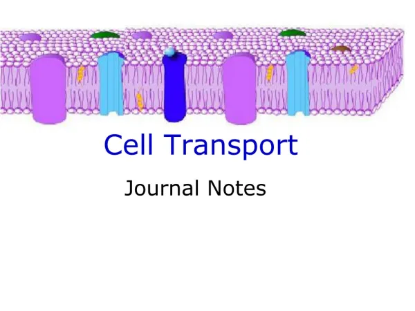
M.Prasad Naidu MSc Medical Biochemistry, Ph.D,. Cell Transport
Synthesis of secretory proteins - review 1. N-terminal signal sequence is synthesized 2. Signal bound by SRP, complex docks with SRP receptor on ER membrane 3. Signal sequence binds to translocon, internal channel opens, inserted into translocon 4. Polypeptide elongates, signal sequence cleaved 5. ER chaperones prevent faulty folding, carbohydrates added to specific residues 6. Ribosomes released, recycle 7. C-terminus of protein drawn into ER lumen, translocon gate shuts, protein assumes final conformation
Synthesis of integral membrane protein • Integral membrane protein may, or may not have N-terminal signal sequence • In absence of N-terminal signal sequence, internal signal sequence bound by SRP • Animation:ERimport.mov • SRP-protein-ribosome complex docks with SRP receptor, C-terminal portion of protein cotranslationally inserted into lumen of ER • Mature protein transverses ER bilayer forming integral membrane protein • NOTE: Orientation of protein within membrane dependent upon cluster of charged residues adjacent to internal signal sequence • In presence of N-terminal signal sequence, integral membrane protein produced by stop-transfer signal that forms transmembrane domain
Secretory Pathway • Once a protein has entered exocytotic pathway, in general, it never returns to cytosol (notable exception is misfolded proteins - retrograde transport for degradation) • In the absence of a sorting signal, protein will follow constitutivesecretory pathway (i.e., directed to plasma membrane) in transport vesicles • Some proteins contain retention signals (e.g., KDEL in C-terminus of some ER proteins)
Secretory Pathway • In specialized cells, regulatedsecretory pathway leads to packaging of product in secretory vesicles
Asymmetry of proteins and lipids maintained during membrane assembly • Orientation of a protein (asymmetry) is determined upon entry into ER, does not change during transit to other membrane/organelle • Fusion of a vesicle with the plasma membrane preserves the orientation of any integral proteins embedded in the vesicle bilayer • Animation: Secretion.mov
GDP GDP GTP GTP GAP Pi Small GTPases Act as Molecular Switches ARF - vesicular transport Ran - nuclear transport Rab - regulated secretion, endocytosis, intracellular transport Rho - formation of actin cytoskeleton Ras - growth and differentiation signaling pathways GEF “Inactive” “Active” GTP exchange for bound GDP, facilitated by Guanine-nucleotide Exchange Factors (GEFs), “activates” protein (usually resulting in conformational change). Hydrolysis of GTP GDP, accelerated by GTPase-Activating Proteins (GAPs), “inactivates” complex.
Intracellular Transport Vesicles Step 1: Coat assembly initiated Step 2: ARF recruits coat proteins Step 3: Vesicle budding Step 4: Coat disassembly Step 5: Vesicle targeting (v-SNARE) Step 6: General fusion machinery assembles (NSF, SNAP) Step 7: Vesicle fusion Step 8: Retrograde transport NOTE: Botulinum B toxin, one of most lethal toxins known (most serious cause of food poisoning), is a protease that cleaves synaptobrevin (one v-SNARE involved in fusion of synaptic vesicles) and inhibits release of acetylcholine at neuromuscular junction. Possibly fatal, depending on dose taken.
Signal sequences target proteins to their correct destinations • Signal sequences identified for cytosolic proteins destined for nucleus, mitochondria, peroxisomes • Animation: Targeting.mov • Nuclear import via nuclear pore complex. Bidirectional transport, accomodates large, complex structures (e.g., ribosomes), nuclear localization signal (NLS) not cleaved during transport. • Mitochondrial (mt) genome encodes 13 proteins, must import remainder. Matrix proteins must pass through outer and inner mt membranes. Proteins must be unfolded by chaperone proteins before translocation. Signal sequence usually cleaved. • Peroxisomes can import intact oligomers (e.g., tetrameric catalase). Zellweger Syndrome - mutation in genes (peroxins) involved in peroxisome biogenesis (or certain peroxisomal enzymes)
Major mechanisms used to transfer material and information across membranes • Cross-membrane movement of small molecules • Diffusion (passive and facilitated) • Active Transport • Cross-membrane movement of large molecules • Endocytosis • Exocytosis • Signal transmission across membranes • Cell surface receptors • 1. Signal transduction (e.g., glucagon cAMP) • 2. Signal internalization (coupled with endocytosis, e.g., LDL receptor) • Movement to intracellular receptors (steroid hormones; a form of diffusion) • Intercellular contact and communication Table 43-11
Passive Mechanisms Move Some Small Molecules Across Membranes • Passive transport down electrochemical gradients by simple or facilitated diffusion • passive diffusion (e.g., gases) limited by concentration gradient across membrane, solubility of solute, thermal agitation of that specific molecule • Active transport, against gradient, requires energy
Ion Channels Selectively Transport Charged Molecules • Specific channels for Na+, K+, Ca2+, and Cl- have been identified • Channels are very selective, in most cases, to only one type of ion • Subset of K+ channels (“K+ leak channels”) open in “resting” cell • make plasma membrane more permeable to K+ than other ions, maintains membrane potential
Activities of Ion Channels Can Be Regulated • Channels are “gated” - open transiently • Ligand-gated channels - specific molecule binds receptor, open channel (e.g., acetylcholine) • Voltage-gated channels - open (or close) in response to changes in membrane potential • Ion channel activities are affected by certain drugs • Mutations in genes encoding ion channels can cause specific diseases (e.g., Cystic fibrosis - mutations in CFTR, a Cl- channel)
Net diffusion of substance depends on: • Its concentration gradient across membrane - solutes move from high to low concentration • Electrical potential across membrane - solutes move toward solution with opposite charge (inside of cell usually has negative charge) • Permeability coefficient of substance • Hydrostatic pressure gradient across membrane - pressure will rate and force of collision with membrane • Temperature - temperature will particle motion and frequency of collisions between particles and membrane
Types of transport systems • Classified by direction of movement and whether one or more unique molecules are moved • Uniport system moves one type of molecule bidirectionally • Cotransport systems transfer one solute dependent upon simultaneous or sequential transfer of another solute • Symport - moves solutes in same direction (e.g., Na+-sugar transporters or Na+-amino acid transporters) • Antiport - moves two molecules in opposite directions (e.g., Na+ in and Ca2+ out)
Transport with carrier proteins • Facilitated diffusion and active transport used to transport molecules that cannot pass freely through lipid bilayer by themselves • Both involve carrier proteins; show specificity for ions, sugars, and amino acids; and resemble a substrate-enzyme reaction (but with no covalent interaction) • But, facilitated diffusion can be bidirectional, while active transport usually unidirectional • And, active transport always against gradient, requires energy • Specific binding site for solute • Carrier is saturable (has maximum rate of transport - Vmax) • There is a binding constant (Km) for the solute, so the whole system has a Km • Structurally similar competitive inhibitors block transport
Facilitated Diffusion • Some solutes diffuse across membranes down electrochemical gradients more rapidly than expected from size, charge, and partition coefficients • “Ping-Pong” mechanism explains facilitated diffusion • Carrier protein exists in two principal conformations: • “Pong” state - exposed to high [solute], solutes bind to specific sites on carrier protein • Conformational change exposes carrier to lower [solute] - “ping” state • Process is reversible, net flux depends on concentration gradient
Facilitated Diffusion • Rate of solute entry into cell determined by: • Concentration gradient across the membrane • Amount of carrier available (key control step) • Rapidity of solute-carrier interaction • Rapidity of conformational change (both loaded and unloaded carrier) • Hormones regulate by changing number of transporters available • e.g., insulin increase glucose transport in fat and muscle by recruiting transporters from intracellular reserve
Active Transport • Transport away from thermodynamic equilibrium • Energy is required (from hydrolysis of ATP, electron movement, or light) • Maintenance of electrochemical gradients in biologic systems consumes ~30-40% of total energy expenditure of cell • Cells, in general, maintain low intracellular [Na+] and high intracellular [K+], with net negative electrical potential inside • Gradients maintained by Na+-K+ ATPase • Ouabain or digitalis (cardiac glycosides used to treat congestive heart failure) inhibits ATPase by binding to extracellular domain. (Raises intracellular [Na+], Na+/Ca2+ antiporter functions less efficiently with lower [Na+] gradient, thus fewer Ca2+ ions exported, intracellular [Ca2+] increases causing muscle to contract more strongly.)
Glucose Transport - Several Mechanisms • In adipocytes and muscle, glucose enters by facilitated diffusion • In intestinal cells, glucose and Na+ bind to different sites on glucose transporter (symport) • Na+ enters cell down electrochemical gradient and “drags” glucose with it • To maintain steep Na+ gradient, Na+-glucose symport depends on low intracellular [Na+] maintained by Na+-K+ pump • A uniport allows glucose accumulated in cell to move across different membrane toward a new equilibrium
Endocytosis • Process by which cells take up large molecules • Source of nutritional elements (e.g., proteins, polynucleotides) • Mechanism for regulating content of certain membrane components (e.g., hormone receptors) • Most endocytotic vesicles fuse with lysosomes • hydrolytic enzymes digest macromolecules (yields amino acids, simple sugars, and nucleotides) • Two general types of endocytosis • Phagocytosis - specialized cells (e.g., macrophages) ingest large particles (viruses, bacteria)
Fluid-phase Receptor-mediated endocytosis endocytosis Endocytosis • Pinocytosis - property of all cells • Fluid-phase pinocytosis - nonselective uptake of a solute by small vesicles • loss of membrane replaced by exocytosis • Absorptive pinocytosis - receptor-mediated selective process • permits selective concentration of ligands from medium, limits uptake of fluid or soluble unbound macromolecules • vesicles derived from coated pits (clathrin) • fate of receptor/ligand depends of particular receptor • e.g., LDL receptor recycled, LDL processed in lysosomes • EGF receptor degraded (receptor downregulation)
Exocytosis • Most cells release macromolecules to the exterior • Signal for regulated exocytosis is often a hormone • binds to cell-surface receptor, induces local and transient change in [Ca2+] that triggers exocytosis • Molecules released by exocytosis fall into 3 categories • Attach to cell surface and become peripheral proteins (e.g., antigens) • Become part of extracellular matrix (e.g., collagen) • Enter extracellular fluid and signal other cells (e.g., insulin)
Membrane proteins classified as: receptors, transporters, ion channels, enzymes, and structural components Member of each class often glycosylated mutations affecting this process may alter function Mutations Affecting Membrane Proteins Cause Diseases