
Mycobacterium Tuberculosis Infections
E N D
Presentation Transcript
Order: Actinomycetails Family: Myobacteriaceae Genus: Mycobacterium
Divided into 4 main groups: • Mycobacterium tuberculosis • M. bovis • Atypical mycobacteria • M. leprae
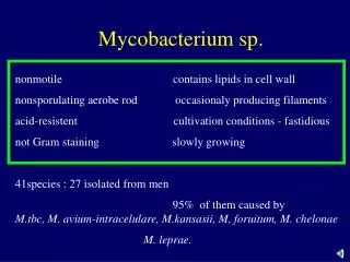
All mycobacteria are intracellular, acid fast bacilli; they are in pairs or in small groups. Sometimes show branching filamentous forms. They are aerobic, non motile, non spore forming non capsulated slow growing forms.
The bacilli appear red in sharp contrast to the blue counter stained background.
Mycobacterium tuberculosis: • They are slender, straight or slightly curved, 2-4 x 0.3-0.5m. in tissues they occur singly or in pairs or in small bundles of parallel bacilli( serpentine cords). While in culture, longer filamentous forms are sometimes seen with swollen or club- shaped forms which indicate the close relation of these organisms to the higher fungi hence they are called mycobacteria(fungus- like bacteria)
if smear is prepared from culture the bacilli appear short, thick and tend to arranged in a sort of bundles (cord formation).
Stained and recognized by acid fast stain with discoloration by 20-25% H2SO4 , or 3% acid + 95%alcohol. They are slow growing with generation time 12-16 hours, therefore 4-6 weeks required for good growth. They do not grow on ordinary media,
they require special media for growth, such as:- • Lowenstein- Jensen medium: which contain egg yolk, glycerol and malachite green. • Glycerol broth: the organism forms a thick, wrinkled, creamy pellicle on top of the medium if undisturbed. Otherwise, will grow as floccules due to the lipid content of the cell wall.
*Cord factor: two mycolic acid molecule with one trehalose. • Organism resistance to acid and alkalis. • Organism resistance to antimycobacterial drugs; strain were found to be resistant to isoniazid (isonicotinic acid hydrazide-INH) and also other drugs due to chromosomal mutation because they don’t posses plasmids.
Resistance and survive if protected from the sun, the bacilli can survive desiccation for weeks or even months and in dried state of sputum or excreta for 6-8 months and in 5% phenol for several hours. The bacilli are killed at 60 C for 15-20 min. and in 2 hours when exposed to sunlight. Variation and virulence and the formation of the variant BacilleCalmette- Guerin”(BCG).
In man, tuberculous infection almost exclusively occurs by means of inhalation of a single virulent tubercle bacillus (T.B.) or, at most, few bacilli in a droplet of sputum, or other infected material. Since no mycobacterium produce exotoxins or endotoxins, they can survive and multiply within a cellular vacuole of the phagosome. They can produce a special protein called “exported repetitive protein” which prevents the fusion of the phagosome with the lysosome and form the phagolysosome, therefore it will escape the killing and degradation by the lysosomal contents and lead to The formation of (Ghon focus or primary focus).
Since the organisms live in the highly oxygenated areas,they will be deposited in bronchoils or the aleveoli, which will be taken by the fixed macrophages where they will start their multiplication intracellularly, and when increase in number, the cell bursts and new phagocytes become infected and repeat the cycle. During this period of 3-4 weeks, sufficient antigen is released to sensitize the host to tuberculoprotien which develops an allergic response.
This lead to an acute inflammatory process followed by central necrosis which appears at the site of the primary focus, which contains large number of organisms. Few bacilli may be carried by phagocyte through the lymphatic channels to the regional lymph nodes, which become enlarged and may caseate. The whole process which is the primary focus , the draining lymph channels, and the enlarged caseous regional lymph nodes produce what is called (the primary complex)
The future course of the case depend on certain factors such as; • The favorable cases, the primary lesion and the involved lymph nodes undergo healing and calcification. • Under unfavorable conditions, progressive lesion may develop and produce miliary tuberculosis, the tuberculous bronchopneumonia, and may involve other organs such as: spleen, liver, brain, kidneys and menings.
The immune response of the human body to the presence of T.B. is indicated by the appearance of agglutinins, precipitins, opsonins and complement fixation antibodies in the serum. These antibodies are found in low titer, and are not used for diagnostic purposes, and none of their antigenic substances are found to be related to virulence, and has no or little effect on the tubercle cells.
Resistance to the organism required by the cell mediated immunity, by T-cell and macrophages. Patient deficient in cellular immunity, such as AIDS patients are at much risk for disseminated tuberculosis. Mutation in the δ- interferon receptor gene is another cause of defective cellular immunity that predispose to severe tuberculosis.
A gene called NRAMP determines the natural resistance to tuberculosis. People who have mutations in this gene have a much higher rate of clinical tuberculosis than those with the normal gene. The NRAMP protein is located in the membrane of the phagosome in macrophages and plays an important role in killing the organism within the phagosome.
Tuberculin test: • Koch’s phenomenon. • Purified protein derivative (PPD) by Seibert. • Tuberculin test is used to investigate infection or previous vaccination with BCG and not necessarily active disease.
The following techniques are employed in tuberculin testing. The Mantoux test: the dose used is 1-5 tuberculin units (each 1.0 t.u. =0.000028 PPD) is injected intracutaneously and as superficially as possible. Usually into the middle- third of the forearm. The positive reaction appears within 1-2 days showing papule with erythema, or a vesicle may develop in the central part of the indurated area and even with necrosis.
2. The von pirquet test: in this test 2 fine scratches are made not to be too superficial and should not bleed. Tuberculin containing a drop of 1% adrenaline/ml is rubbed into the scratched area with a glass rod and left to dry for 5 min. positive reaction appear after 72 hours showing an induration of not more than 5 mm. as determined by palpation.
3. The Heaf multiple puncture test: this test requires a special multiple puncture apparatus designed to release 6 needles automatically when the end plate is pressed vertically on the skin.
False positive results: • Histoplasmacapsulatum infection • A hot platinum needle is used • Formation of a small hematoma • From a slight skin infection at the site of injection • From bacterial contaminated tuberculin
False negative results: • Old deteriorated tuberculin • Deeper injection • Leakage of tuberculin after injection • Very severe infection such as miliary tuberculosis • During cortisone administration • During measles infection ( lymphocytes become non susceptible to stimulating effect of PPD ) • Malignant lymphomas • Sarcoidosis.
Clinical manifestations and chest X-Ray are not enough to give the clear diagnosis without a confirmatory laboratory tests. Specimens expected to reach the laboratory for acid fast bacilli(AFB) are sputum, urine, CSF, gastric washings, pleural fluid, infected tissues, laryngeal swab, feces and pus.
The laboratory tests may include: • Direct microscopic examination by light and fluorescent microscope; by using the acid fast stain and also using the auramine dye. • Concentration method: by using the NaOH to kill and dissolve all the mucous materials and the bacterial and epithelial cells without affecting the T.B. cells. Then the specimen centrifuged. • Cultural method: by growing the concentrated specimen on the Lowenstein-Jensen medium, or on liquid medium. • Animal inoculation.
M. Tuberculosis is differentiated from other mycobacterium by its production of the niacin and also produces catalase.
Treatment and resistance: Since the treatment last very long (6-9 months), new resistant strains may emerge. Therefore a multidrug therapy is used such as: isoniazid (INH) + Rifampin + Pyrazinamide. Pyrazinamide is given for two months and the other two are continued for at least 6 months. In immune compromised patients (such as AIDS). Or with disseminated disease, or with INH resistant strains; a fourth drug is added which is ethambutal, and the fourth drug may be given for 9-12 months. Although the treatment lasts for months, the patient sputum became non infectious within 2-3weeks.
INH resistant strain could be detected by using the luciferase enzyme assay; this enzyme is produced by the firefly which produces flashes of light by the help of ATP. If the organism is resistant will produce ATP and hence gives light. If it is sensitive, then it will be damaged and much less light will be produced.
Atypical mycobacteria: This group of microorganisms resembles M. tuberculosis in certain aspects and differs from it in other aspects. Such as their non pathogenicity to guinea pigs and their spread in the environment. They are classified into four groups according to their rate of growth and pigment production:
Group 1 (photochromagens) produce yellow-orange pigment in light, e.g. M. kansasii (cause lung disease) and M. marinum (cause skin disease). • Group2 (schotochromagens) produce the pigment in dark, e.g. M. scrofulaceum (cause granulomatous cervical adenitis). • Group 3( Nonchromagens) no pigment produced, e.g. M. avium and M. intracellular (produce pulmonary disease in immunocompromised)
Group 4 (Rapid growing Mycobacteria) grow very fast and produce colonies in 7 days, e.g. M. fortuitum and M. chelonei (rarely cause disease or may be in immunocompromised patients) M.Abscessus ( cause chronic lung infections) and M. smegmatis ( non pathogenic) are also rapidly growing mycobacteria.
M. leprae • It is an ancient disease. • Cell characters are almost similar to that of T.B. • They are intracellular occurs singly or in large numbers lying parallel to each other arranged in bundles or clusters like “match stick pack” or “packet of cigars”. This arrangement is due to lipid like substance” the globia”
The live bacilli are solid and uniformly stained, while the dead cells are fragmented and granular, and depending on this character; the effect of drug could be detected. They are non motile, non spore forming and non capsulated. Humans are the natural hosts. The optimum temp. is 300 C, therefore grows on face and superficial nerves. The organism has never been cultivated on laboratory media. It can be grown only in the footpad of the mouse or the armadillo.
The generation time is between 14-18 days. • It is only mycobacteria that produce the enzyme phenolase which converts the compound 3,4 dihydroxyphenelanine to color product. • Gives positive skin test with lepromin prepared from lepromatous nodule and equally well to PPD or to tuberculin prepared from M.pheli.
Pathogenesis:- • It is an infectious disease, but infections are acquired by prolonged contact with the lepromatous leprosy. • Do not necrose or caseate like tuberculosis, but frequently ulcerate with discharge of infectious exudates. • There are two forms of leprosy, 1- lepromatous and 2- tuberculoid :
In lepromatous leprosy: the bacilli are easily demonstrated by acid fast stain of skin lesions or nasal scrapings. The lipid laden macrophages called “foam cells” containing many acid fast bacilli are also seen in the skin. In tuberculoid leprosy: very few organisms are seen and the appearance of typical granulomas is sufficient for diagnosis.
Treatment: Dapsone (diaminophenylsulfone) + Rifamcin + clofazimine Dapsone used to be given alone, but due to the emergence of resistant strains, now is given together with these two drugs for at least 2 years.
Actinomycete • These are true bacteria (related to corynebacteria and mycobacteria), but they form long branching filaments that resemble hyphae of fungi. • They are Gram +ve but some (such as Nocardia asteroids) are weakly acid fast.
Actinomyces Israeli • Gram positive non spore forming branching bacilli • Anaerobic, grow best with 5-10% CO2 • All species are found as normal commensally in the oral cavity; some are isolated from deep caries. Other from brain abscess.
Pathogenesis The disease is characterized by multiple abscesses and granulomata, tissue distraction, extensive fibrosis, and the formation of sinuses.
Infected tissue show large mycelia like formation which are embedded in an amphorous protein- polysaccharides matrix and surrounded by a zone of bacterial products, which is Gram negative, club like structures, which sometimes become visible to the naked eye and look yellow in appearance called (sulfur granules).
These sulfur granules may become dark brown and very hard due to deposition of calcium phosphate. • The most affected area are the cervicofacial and the jaws • The disease is mainly endogenous as a result of dental caries or dental extraction.
Sometimes, thoracic (such as the lungs), or abdominal such (as the appendix and colon) actinomycosis may occur as a result of aspiration from the mouth, leading to the formation of sinuses on the chest and abdomen respectively.
Laboratory diagnosis: Specimen obtained from lesion by aspiration or biopsy is stained by Gram stain and weak acid fast stain to observe the mycelia- like formation and the sulfur granules respectively. • Culture on brain heart infusion or blood agar show spider like colonies. • No serological tests are used.
Treatment • The problem is the penetration of the drug to the fibrotic tissues and reaching the organism which may lead to recurrent infection. • The drug of choice is penicillin G and ampicillin coupled with surgical drainage.