Pregnancy: From Detection to Development
Discover the intricacies of pregnancy, from detecting it with tests like urine and blood tests to understanding factors influencing fetal growth and gestation. Explore the formation of the fetus and the physical changes during pregnancy. Learn about multiple pregnancies, ultrasound scans, fetal development stages, and factors affecting growth. Explore how genetics, environment, and hormones impact the growth of the baby.


Pregnancy: From Detection to Development
E N D
Presentation Transcript
Topics to be covered • What is pregnancy? • How to detect pregnancy • Fetus formation • Factors affecting fetal growth • Factors affecting gestation • Physical changes during pregnancy
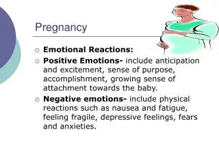
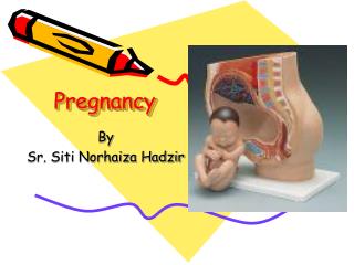
What is pregnancy? • Pregnancy is the development of an embryo and subsequently a fetus in the uterus of a woman for a length of time • During this stage, physical changes occur to the woman • Pregnancy is maintained by hormones (HCG and Progesterone)
Multiple pregnancy • A pregnancy with two or more fetuses. Twins - 2 fetuses, Triplets - 3 fetuses, Quadruplets - 4 fetuses, Quintuplets - 5 fetuses, Sextuplets - 6 fetuses and Septuplets - 7 fetuses • Naturally occurring factors causing multiple pregnancy are: • heredity- A family history of multiple pregnancy increases the chances of having twins • older age- Women over 30 have a greater chance of multiple conception. • high parity-Having one or more previous pregnancies, especially a multiple pregnancy, increases the chances of having multiples. • race-African-American women are more likely to have twins than any other race. Asian and Native Americans have the lowest twinning rates. Caucasian women, especially those over age 35, have the highest rate of higher-order multiple births (triplets or more).
How to detect pregnancy? • Urine test – detect hCG • Blood test – detect hCG • Ultrasound • Milk test – P4 (animals) • Blood test – PMSG (animals)
Urine test • The hCG Urine Pregnancy Test Strip is a test kit based on a visual, qualitative principle for the determination of Human Chorionic Gonadotropin (a glycoprotein hormone secreted by the developing placenta after fertilization) in urine specimens. • Pregnancy Test Strips are over 99% accurate and are capable of detecting 20mIU/ml/hCG. Can test accurately 6 to 8 days after conceiving - and 7 days after missed period. • Appearance of hCG soon after conception and its subsequent rise in concentration during early gestational growth make it an excellent marker for the early detection of pregnancy
hCG (Human Chorionic Gonadotrophin) • The developing placenta begins releasing hCG into blood as early as 6 days after implantation. Some hCG also gets passed in the urine • HCG helps to maintain pregnancy and affects the development of fetus • Levels of hCG increase steadily in the first 14 to 16 weeks following LMP, peak around the 14th week following LMP, and then decrease gradually • The amount that hCG increases early in pregnancy can give information about pregnancy and the health of the baby. Shortly after delivery, hCG can no longer be found in the blood • More hCG is released in a multiple pregnancy, such as twins or triplets, than in a single pregnancy • Less hCG is released if the fertilized egg implants in a place other than the uterus, such as in a fallopian tube. This is called an ectopic pregnancy.
Ultrasound • An ultrasound test is a radiology technique, which uses high- frequency sound waves to produce images of the organs and structures of the body. It involve no radiation and studies have not revealed any adverse effects. • The sound waves are sent through body tissues with a device called a transducer placed directly on top of the skin, which has a gel applied to the surface. • The sound waves that are sent by the transducer through the body are then reflected by internal structures as "echoes." which return to the transducer and are transmitted electrically onto a viewing monitor.
Ultrasound pictures Week 5 Week 8 Week 11 Twins Twins kicking
Fetus formation • Gene dependant • Size dependant on nutrition and health of mother, parity (primiparous mothers have small babies as compared to multiparous mothers), mother’s size, pregnant more than one baby and self-damage caused by smoking, drug addiction, alcoholism etc • Small sized baby is due to prematurity or even if full-term, there must be a factor to cause a retarded growth for the baby
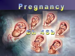
Fetal Development • Heart and brain develop from 3rd week • Heart starts to pump blood from week 4-5 • Feet and hands starts to develop and tail at coccxy starts to shrink • Embryo is less than an inch long at week 5 • Hands and feet is visible and nose also starts to form • At week eight, it is about an inch long • By week 9, embryo is called a fetus • Sexual organs starts to form but sex is not yet determine • Other organs also starts to form and develop until birth
Rate of fetal growth is slow until week 20 but accelerate to a maximum at week 30-36 • Peak of growth velocity is on week 8 • Fetal nutrition is from CHO (glucose), amino acids and lactate. Fatty acids, vitamins and minerals are also transferred to the fetus via the placenta
Factors affecting fetal growth • Genetic (species, breed, genotype) • Environmental (nutrition, size, parity, size and blood circulation of placenta) • Fetal hormones (thyroid, growth hormone, somatomedins)
Gestation length: 280 days or 40 weeks or 9 months and 10 days • LMP – Last Menstrual Period • EDD – Estimated Delivery Date (First day of last menstrual period plus 280 days) • Trimester – 3 months • Human – 3 trimesters
LMP – 31/1/2009 • Menstrual cycle – 28 days • Add 40 weeks or 9 months and 10 days (280 days) • EDD – 7/11/2009 (An estimation!)
Factors affecting gestation length • Maternal factor – age of mother • Fetal factor – number of fetuses, gender, adrenal and pituitary function • Genetic – species, breed, fetal genotype • Environmental factors – nutrition, temperature, season
Physical changes during pregnancy • No menstruation • Nausea in first trimester • Back and hip pains • Increase in body weight • Pigmentation of skin especially in fair-skinned women (cholasma – mask of pregnancy) especially at the facial region • ‘Quickening’ or baby movements in uterus – occurs at 5 months pregnancy onwards • ‘Braxton-Hicks contraction at 6-7 months pregnancy • ‘Engagement’ at 8 months pregnancy • Others eg pica (Pica is a pattern of eating non-food materials such as dirt or paper and should last at least 1 month to fit the diagnosis of pica.)
Abnormal pregnancies – Ectopic pregnancy • Occurs when a fertilized egg attaches somewhere other than in the uterus, usually in a fallopian tube (tubal pregnancy). • Because an ectopic pregnancy can cause life-threatening complications, the pregnancy must be ended with medicine or surgery. • An ectopic pregnancy, especially a tubal pregnancy, can be dangerous because the fallopian tube does not stretch as the fertilized egg grows. If a tubal pregnancy is not detected and treated early, the tube may burst. This can be a life-threatening situation and requires emergency surgery. • Pelvic inflammatory disease or tubal surgery increases the risk of having an ectopic or tubal pregnancy by creating scar tissue that may block the fallopian tube.
Abnormal pregnancies – Molar pregnancy • A mass of abnormal tissue (hydatidiform mole) that comes from the placenta inside the uterus, which triggers symptoms of pregnancy. About 1 : 1,000 women with early pregnancy symptoms has a molar pregnancy. Two types of molar pregnancy: complete and partial. • Complete molar pregnancy. In place of a normal placenta/embryo, the hydatidiform mole is abnormal placental tissue that grows into a grapelike cluster that can fill the uterus. • Partial molar pregnancy. The placenta grows abnormally into molar tissue. Any fetal tissue that develops is likely to have severe defects.