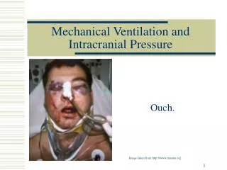
INTRACRANIAL PRESSURE
200 likes | 460 Vues
INTRACRANIAL PRESSURE. JULY 2004. CEREBRAL BLOOD FLOW. CBF affects cerebral volume, oxygen consumption and removal of waste products. Brain receives 15-20 % of cardiac output. Grey > than white matter.

INTRACRANIAL PRESSURE
E N D
Presentation Transcript
INTRACRANIAL PRESSURE JULY 2004
CEREBRAL BLOOD FLOW • CBF affects cerebral volume, oxygen consumption and removal of waste products. • Brain receives 15-20 % of cardiac output. Grey > than white matter. • Receives blood via carotid arteries (80%) and vertebrobasilar arteries (20%). Form Circle of Willis • Venous blood travels in cerebral veins in arachnoid mater before draining into dural venous sinuses.
AUTOREGULATION • In the healthy brain CBF remains constant despite changes in arterial pressure ie. constant between MAPs of 50-150mmHg • In chronic hypertension this is shitfted to the right.
REGULATION OF CBF • CBF increases with metabolism (highest CBF occurs during seizures) • Carbon dioxide- potent vasodiltator. Increasing PaCO2 by 1kPa increases CBF by 30%. At PaCO2 >10 or <4kPa these changes are less marked • In normal range oxygen has little effect on CBF but once PaO2 is <7kPa it causes vasodilatation • Temperature. Hypothermia reduces CBF • Blood viscosity • Anaestheticagents • Neurogenic control – cerebral vessels have supply from parasympathetic and sympathetic nerves
BLOOD BRAIN BARRIER • Semi-permeable capillary membrane • Regulates the movement of all solutes and hence water into CNS extracellular space and so governs cerebral volume • With trauma or inflammation the BBB becomes incompetent and places brain at risk of swelling
CEREBROSPINAL FLUID • Clear aqueous fluid bathing CNS providing support and protection from trauma • Produced at rate of 500-600ml a day • Turnover time 4-5 hours so total volume is 140ml • Formed in choroid plexus in the lateral ventricles and flows into subarachnoid space around brain and spinal cord • 80-90% is absorbed at cranial site through arachnoid villi. Rest absorbed at spinal sites.
INTRACRANIAL PRESSURE • Determined by the relationship between rigid cranium and volume of intacranial contents • Intracranial contents – • Brain tissue 85% • CSF 10% • Blood 5% • Pathological circumstances can produce 4th compartment –the mass lesion
INTRACRANIAL HYPERTENSION • Defined as sustained ICP >15 mmHg • Blood and CSF communicate with extracranial compartments and allow a degree of adaptability • Three compensatory mechanisms- • Translocation of intracranial CSF through foramen magnum to subarachnoid space around spinal cord • Increased CSF absorption by arachnoid villi • Translocation of blood out of cranium • Once these methods are exhausted there is an abrupt rise in ICP with small increase in cerebral volume • Cerebral compliance and capacitance
CAUSES OF RAISED ICP • Increased brain bulk- space occupying lesion, cerebral oedema • Increased CSF volume – benign intracranial hypertension, hydrocephalus • Increased cerebral blood flow • Increased arterial blood - hypoxia, hypercarbia, halogenated inhalational agents • Increased venous blood –coughing, straining, increased intrathoracic or intra-abdominal pressure,obstruction to venous drainage in neck, head down position
CEREBRAL PERFUSION PRESSURE • CPP = MAP – ICP • IF CPP is <70mmHg, it is associated with loss of cerebral autoregulation and may increase secondary insults to the brain • In management of patient with a head injury the strategy is to maintain CPP > 70 mmHg
CEREBRAL OEDEMA • Cerebral oedema commonly accompanies neuropathological processes. It is defined as an increase in water content of brain • 3 forms • Vasogenic –failure of capillary permeability when BBB is disrupted • Cytotoxic –failure of NaK pump in brain cells due to ischaemia leads to accumulation of Na and water in these cells • Interstitial –in hydrocephalus the pressure in ventricles may cause CSF to leak into ECF • In clinical practice it is usually due to vasogenic and cytotoxic processes
CONING • Unrelenting cerebral swelling and increased ICP will ultimately lead to brain displacement • Herniation typically takes place in following sites • Cingulate – one hemisphere is pushed under falx cerebri compressing the ant. cerebral artery • Temporal – the medial temporal lobe is pushed through the tentorium • Tonsillar – the brainstem is pushed through the foramen magnum
MEASURING ICP • Supratentorial – Epidural, subdural, intraventricular, intraparenchymal • Involve insertion of a catheter with zeroing at level of ext. auditory meatus • Ventricular is gold standard – also allows drainage of CSF to reduce ICP • ALSO infratentorial and lumbar techniques • Indications for ICP monitoring – • GCS<9 and abnormal CT • GCS<9 and normal CT but >40, low BP or motor posturing
TREATMENT OF RAISED ICP • ABC • Maintenance of cerebral perfusion may require invasive haemodynamic monitoring +/- ICP monitoring • Adequate oxygenation • Ventilation to hypocarbia or normocarbia ( aggressive hyperventilation can cause vasoconstriction) • Maintenances of MAP to maintain CPP • Head elevation • Prevention of venous obstruction – keep head neutral • Adequate sedation or paralysis to prevent coughing
SPECIFIC MEASURES • Surgical intervention • Mannitol – reduces CSF formation, lowers blood viscosity and reduces brain vol. • Frusemide to give rapid diuresis • Steroids to reduce vasogenic oedema around masses • Other cerebroprotective agents – thiopentone • Mild to moderate hypothermia to reduce metabolic rate • Tight glycaemic control • Seizure control