Intracranial Pressure Concepts
290 likes | 1.13k Vues
Intracranial Pressure Concepts. Michelle Hill RN, BSN, CNRN, CCRN, SCRN Clinical Nurse Educator Neurocritical Care. Review intracranial pressure concepts (ICP) Discuss cerebral hemodynamics Discuss herniation syndromes Discuss management of increased ICP

Intracranial Pressure Concepts
E N D
Presentation Transcript
Intracranial Pressure Concepts Michelle Hill RN, BSN, CNRN, CCRN, SCRN Clinical Nurse Educator Neurocritical Care
Review intracranial pressure concepts (ICP) • Discuss cerebral hemodynamics • Discuss herniation syndromes • Discuss management of increased ICP • Discuss types of ICP monitoring devices Objectives
Intracranial pressure is the pressure exerted by the intracranial contents of brain tissue, blood, and cerebrospinal fluid (CSF) within the skull. • Fluctuates within a normal range. • Normal ICP = 0 – 15 mmHg • Moderate elevation ICP = 15 – 40 mmHg • Severe elevation ICP > 40 mmHg • Intracranial hypertension: • ICP >20mmHg for >5 minutes Intracranial Pressure (ICP)
Monroe-Kellie Doctrine • Used to explain why ICP exists • Skull is a rigid, non-distendable box containing 3 volume components: • 80 % brain tissue • 10% blood • 10% CSF • As long as these volumes remain the same, the pressure within the box is unchanged
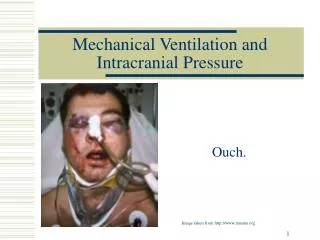
Signs and Symptoms of Increased ICP • Headache (Worse in morning) • Vomiting without nausea • Change in LOC • Change or loss of motor/sensory function • Pupillary changes • Respiratory changes • Papilloedema • Cushing’s Response • Increased systolic blood pressure • Widened pulse pressure • Bradycardia
Cheyne-Strokes • Apneustic • Hyperventilation Respiratory Signs and Symptoms
Progressive deterioration in LOC • Caudal displacement of the diencephalon and midbrain • Pupillary dilitation, B/L ptosis, impaired upward gaze • Extension to pain • Respiratory irregularity Herniation or ICP?
Space-occupying masses • Abscesses • Tumors • Aneurysms • Trauma-hematoma • Cerebral Edema • Vasogenic (extracellular) • Cytotoxic (intracellular) What Causes Increased ICP-Brain
Stroke • Trauma • Conditions that increase blood flow • HTN • PaCO2 • Anesthetic agents • Decreased venous return • HOB flat • Trach ties • Neck flexion More causes of Increased ICP-Blood
Increases in CSF volume • Obstruction of CSF pathways • Non-Communicating hydrocephalus • Decreased CSF absorption • Communicating hydrocephalus • Subarachnoid hemorrhage • Overproduction of CSF • Choroid plexus papillomas More Causes of Increased ICP-CSF
Required to provide oxygenation to the brain tissue • Approximate CBF is 55mL/100g of brain tissue per minute • 450-1000mL/min to the whole brain • Brain receives 20% of total cardiac output and uses 20% of oxygen consumed in the basal state. Cerebral Blood Flow (CBF)
Autoregulation • Ability of an organ to maintain a constant blood flow • Major homeostatic and protective mechanism • Provides a constant CBF by adjusting the diameter of blood vessels. Cerebral Blood Flow Regulation
Arterial carbon dioxide pressure affects the CBF by affecting the arterioles of the brain. • PaCO2 > 45 mmHg causes inappropriate vasodilation of the arterioles which ↑ CBF. • PaCO2 < 35 mmHg causes constriction of the arterioles which ↓ CBF. • PaO2 <50 mmHg also causes cerebral vasodilation. Cerebral Blood Flow
CPP is the blood pressure gradient across the brain • CPP is the difference between the mean arterial pressure (MAP) and the intracranial pressure (ICP) • Any blood coming into the brain must overcome the ICP to enter the intracranial contents and perfuse brain cells. Cerebral Perfusion Pressure (CPP)
CPP = MAP – ICP • More important than ICP value • Normal CPP range is 70 – 100 mmHg • CPP < 60 = ischemia • CPP < 40 = infarct • CPP – 0 = brain death Cerebral Perfusion Pressure
These are protective mechanisms to assure that the brain is receiving adequate perfusion • If one of the intracranial volumes increases another must decrease to avoid increase in ICP • CSF • Blood • Tissue Compensatory Mechanisms
Cerebrospinal Fluid Component • Displacement of CSF into the spinal subarachnoid space • Decreased production of CSF Compensatory Mechanisms-CSF
Blood component • Vasoconstriction of the blood vessels of cerebral structures (carbon dioxide) • Decrease in the intracranial blood volume • Increased venous outflow • Corrected with positioning Compensatory Mechanisms-Blood
Compensatory Mechanisms-Brain • Brain Tissue Component • Supratentorial • Subfalcine (1) • Uncal (2) • Loss of consciousness • Ipsilateral pupil dilation • Contralateral hemiparesis • Infratentorial (3)
Success of compensatory mechanisms is dependent upon several factors: • Rate of expansion of the volume causing increased ICP • Compliance of the brain • Location of the expanding volume Compensatory Mechanisms
Elevated BP • Ischemia in Medullary vasomotor center-increase in systemic arterial pressure • Intraluminal blood pressure must be higher than the ICP for continued blood flow • Widened pulse pressure • Elevated BP increases CO • Bradycardia • Pressure on the Vagal control in the Medulla • Becomes decreased but bounding to pump blood upward Cushing’s Response
Basic measures • ICP monitor • Mannitol • Hyperventilate Management of ICP
Management of ICP • Craniectomy: excision of a portion of the skull without replacement • Skull bone can be stored in the patient’s abdomen • Considered a life-saving measure for maximal cerebral swelling
It is the complete and irreversible cessation of all brain function • Absence of brain function and all brain stem reflexes • Cerebral blood flow is 0 in brain death • Brain death is the legal definition of death • Spinal reflexes may still be present • Brain Death Protocol Brain Death
References Dunn, L. (2002). Raised Intracranial Pressure. Journal of Neurology, Neurosurgery and Psychiatry. 73 (suppl 1). i23-i27. Germon, K. (1988). Interpretation of ICP pulse waves to determine intracerebral compliance. Journal of Neuroscience Nursing, 20, 344–351. Hickey, J. V. (2009). The Clinical Practiceof Neurological and Neurosurgical Nursing (6th ed.). Philadelphia: Lippincott. March, K. (2004). Intracranial Pressure Concepts and Cerebral Blood Flow. In M. K. Bader & L. R. Littlejohns, AANN Core Curriculum for Neuroscience Nursing (4th ed., pp. 87–114). Philadelphia: Saunders. Slazinski, T., Anderson, T., Cattell, E., Eigsti, J., Heimsoth, S., Holleman, J. & et.al. (2011). Care of the patient undergoing intracranial pressure monitoring/external ventricular drainage or lumbar drainage. American Association of Neuroscience Nurses Clinical Practice Guideline Series. Stevens, R., Huff, J., Duckworth, J., Papangelou, A., Weingert, S. & Smith, W., (2012). Emergency Neurological Life Support: Intracranial Hypertension and Herniation. Neurocritical Care. DOI: 10.1007/s12028-012-9754-5