GASTROINTESTINAL IMAGING
GASTROINTESTINAL IMAGING. MII - Lecture Dr. Janette Fulton 2/2008. GI IMAGING. Single AP radiograph Showing filling of the stomach and proximal small bowel without mass, obstruction or filling defect. AP SUPINE ABDOMEN XRAY GAS PATTERN. STOMACH. COLON. SM. BOWEL.

GASTROINTESTINAL IMAGING
E N D
Presentation Transcript
GASTROINTESTINAL IMAGING MII - Lecture Dr. Janette Fulton 2/2008
GI IMAGING Single AP radiograph Showing filling of the stomach and proximal small bowel without mass, obstruction or filling defect.
AP SUPINE ABDOMEN XRAY GAS PATTERN STOMACH COLON SM. BOWEL Normal abdominal gas pattern with air in the stomach and scattered non-distended loops of large and small bowel.
ESOPHAGEAL DISEASE • ESOPHAGEAL CANCER • HIATAL HERNIA • VARICES • CANDIDA • MALLORY WEISS TEAR • BOERHAAVE’S SYNDROME • ACHALASIA SIGNS / SYMPTOMS • CHEST PAIN • DIFFICULTY SWALLOWING • HOARSENESS
NORMAL ESOPHAGUS Normal double contrast esophagram (barium coating and air distention) Effervescent granules release air with ingestion.
RADIOLOGY DIAGRAM Pathology image X ray image
ESOPHAGEAL CANCER Typical squamous cell carcinoma Poor prognosis from local extension into critical mediastinal structures. (esophagus lacks a serosa) .
ESOPHAGEAL CANCER Distil malignancy may be adenocarcinoma due to Barrett’s esophagus - dysplastic change caused by chronic reflux of gastric contents.
ESOPHAGEAL VARICES Linear tubular filling defects represent distended veins from shunting due to cirrhosis and portal hypertension
CANDIDA ESOPHAGITIS Extensive nodular filling defects in the esophagus in an immunocompromised patient are typical for candida esophagitis.
ACHALASIA Distended esophagus with distil stricture due to Achalasia - Failure of distil sphincter to relax – causing obstruction. Etiology is unknown. Stricture due to cancer or reflux caused scarring have to be considered first. Barium filled esophagus
MALLORY-WEISS TEAR Esophagus shows a linear tear of mucosa of distil esophagus due to vomiting with barium tracking into the wall. Full thickness tear or rupture (Boerhaave’s syndrome) can lead to mediastinitis and death.
HIATAL HERNIA NORMAL ESOPHAGUS *Note distended distil esophagus with herniation of gastric fundus into chest through esophageal hiatus. DIAPHRAGM DIAPHRAGM
HIATAL HERNIA L CXR FINDINGS Mass on chest X- ray posterior to heart may be a large hiatal hernia.
ASPIRATION NORMAL SWALLOW Contrast tracks anteriorly into trachea with aspiration.
GASTRIC DISEASE • ULCER • CANCER • PYLORIC STENOSIS SIGNS / SYMPTOMS • PAIN • ANEMIA • HEMATEMESIS / MELENA • EMESIS • WEIGHT LOSS
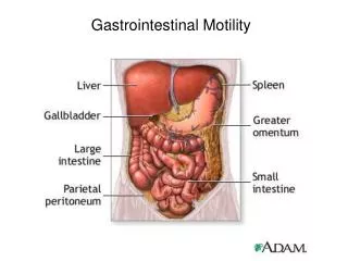
FUNDUS NORMAL GASTRIC ANATOMY DUODENUM ANTRUM BODY JEJUNUM Single AP radiograph showing filling of distil esophagus, stomach and proximal small bowel without mass, obstruction or filling defect. C-LOOP
GASTRIC ULCER Barium collects in ulcer crater Endoscopic view of ulcer
NORMAL GAS PATTERN AIR UNDER THE DIAPHRAGM Perforation of GI tract leads to pneumoperitoneum collecting subdiaphragmaticly on upright x-ray
UPRIGHT ERECT AND DECUBITUS ABDOMEN FILMS SHOW FREE AIR UNDER THE DIAPHRAGM. DECUBITUS Left lateral decubitus (left side dependent) shows air along liver margin. This is the preferred x-ray if the patient cannot stand.
GASTRIC CARCINOMA Note: Narrowed lumen of gastric antrum by infiltrating carcinoma-typical adenocarcinoma – Linitis Plastica
PYLORIC STENOSIS Normal stomach OBLIQUE VIEW Air filled fundus Air filled fundus Barium filled antrum Duodenal bulb Duodenal bulb Barium filled antrum Narrowed pyloric channel Pyloric Stenosis is seen in newborns within the first months. There is a 4:1 male ratio and is due to hypertrophied musculature at the pylorus.
PYLORIC STENOSIS Thickened pylorus Narrowed pyloric channel Narrowed pyloric channel ULTRASOUND
SMALL BOWEL DISEASE • ULCER • OBSTRUCTION • POST-OPERATIVE ILEUS • CROHN’S DISEASE SIGNS / SYMPTOMS • PAIN • HEMATEMESIS • DISTENTION • DIARRHEA
DUODENAL ULCER Note barium collection Centrally with surrounding Edema.
NORMAL SMALL BOWEL IMMEDIATELY AFTER INGESTION OF BARIUM ONE HOUR LATER Early contrast is predominantly in jejunum and later predominately in ileum. (note difference in mucosal fold pattern)
SMALL BOWEL OBSTRUCTION Ng tube ERECT Note dilated small bowel centrally placed with air/fluid levels on upright exam.
PARTIAL SMALL BOWEL OBSTRUCTION DILATED BOWEL * OBSTRUCTION NON DILATED BOWEL Proximal loops are dilated and distil loops are collapsed indicating an obstruction.
CT- SMALL BOWEL OBSTRUCTION PROXIMAL DILATED BOWEL Proximal loops are dilated and distil loops are collapsed indicating an obstruction. Obstruction most likely due to adhesions in a patient with history of abdominal surgery DISTAL NORMAL BOWEL
SM. BOWEL BARIUM STUDY HERNIA CT Note hernia in right lower quadrant on both exams accounting for obstruction. Hernia is likely cause if there is no history of prior surgery.
POST – OP ADYNAMIC ILEUS COLON LARGE AND SMALL BOWEL SM. BOWEL Symmetric dilation of large and small bowel is seen normally as a post operative ileus.
POST – OP ADYNAMIC ILEUS sutures Colon resection
CHROHN’S DISEASE normal Narrowed distil ileum due to chronic inflammation is typical for Crohn’s disease.
COLON DISEASE • APPENDICITIS / DIVERTICULITIS • POLYP / CANCER • VOLVULUS • NECROTIZING ENTEROCOLITIS • GI HEMORRHAGE • SIGNS / SYMPTOMS • RIGHT / LEFT LOWER QUADRANT PAIN • FEVER / ELEVATED WBC’s • DISTENSION / OBSTRUCTION • WEIGHT LOSS • HEMOCULT POSITIVE STOOL / ANEMIA • MELENA / HEMATOCHEZIA
APPENDICOLITH Occasionally a calculus (appendicolith) is seen as the source of appendicitis due to obstruciton of the appendix and inflammation.
ACUTE APPENDICITIS NORMAL DISTENDED APPENDIX WITH LOCAL INFLAMATION.
ABSCESS Catheter has been placed by radiologist using CT guidance draining abscess collection DRAINAGE
SPLENIC FLEXURE NORMAL COLON HEPATIC FLEXURE TRANSVERSE COLON DESENDINGCOLON ASCENDING COLON Normal air contrast barium enema (double contrast-air and barium per rectum) shows filling of colon with air and barium retrograde to the cecum with reflux into the terminal illeum TERMINAL ILEUM CECUM
PEDUNCULATED COLON POLYP (DESCENDING COLON) stalk on polyp--pedunculated
COLON POLYP Polyp on wall without stalk is coated and outlined by barium
COLON OBSTRUCTION Distension extends to distil descending colon.
COLON CANCER Barium enema showing apple-core type constricting lesion with proximal dilation of colon—”APPLE -CORE” constricting lesion
COLON SIGMOID VOLVULUS Dilated horse-shoe shaped sigmoid colon due to volvulus. “COFFEE BEAN SIGN”
COLONVOLVULUS “BEAK SIGN” Barium fills to point of obstruction and twist of sigmoid colon
NECROTIZING ENTEROCOLITIS #1 #2 Air in bowel wall is due to Necrotizing Enterocolits. #1- an infectious complication of premature infants. Air has tracked into the Portal vein and is seen in #2.
DIVERTICULOSIS Barium extends from lumen outward into diverticulum.
DIVERTICULITIS Extensive inflammation, wall thickening and spasm can simulate carcinoma with colonoscopy required to confirm.