Download
1 / 15
150 likes | 238 Vues
Learn about the importance of different thorax views in small animal radiology, including lateral, ventrodorsal, and dorsoventral views. Each view offers unique insights into the thoracic cavity, aiding in the diagnosis of conditions like diaphragmatic hernia. Find out how to position the animal correctly for optimal imaging results.

E N D
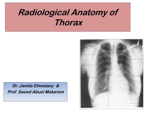
Thorax • Lateral View • Sometimes do both lateral views. • Patient is placed in lateral recumbency with front legs extended cranially. • Head is extended slightly. • View should include entire thoracic cavity • View should be taken at peak of inspiration.
Thorax • Ventrodorsal View with Horizontal Beam • Used to confirm quantitative thoracic fluid or air. • Animal is in lateral recumbency on top of a foam pad. This allows visualization of both sides of thorax. • Forelimbs and head are extended cranially. • Thorax is centered on the cassette. • View includes entire thorax. • Can also be used for the abdomen.
Thorax • Dorsoventral View • Preferred for evaluation of heart because heart is closure to sternum and is in the near-normal suspended position within the thorax. • May be difficult to position larger dogs due to deep chest. • Patient is in sternal recumbency with thoracic vertebrae superimposed over the sternum. • Forelegs are pulled forward, rear legs are in natural crouched position. • Head is placed between two forelimbs • View should include entire thorax, which is all of the ribs. • Exposure should be taken at peak of inspiration to allow complete radiographic visualization of the lung tissue.