Download
1 / 78
780 likes | 896 Vues
This comprehensive overview details the structures and divisions of the respiratory system, including the upper tract (nasal cavity, nasopharynx) and lower tract (larynx, trachea, bronchi). It explores the histological features such as the mucosal layers, glandular components, and muscle arrangement within the trachea and bronchi. Additionally, it discusses surgical procedures like coniotomy and tracheotomy, along with their indications and potential complications. Understanding the intricate anatomy is crucial for safe and effective interventions in respiratory emergencies.

E N D
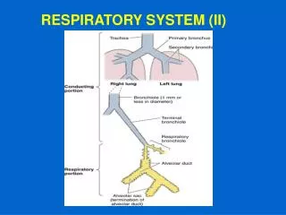
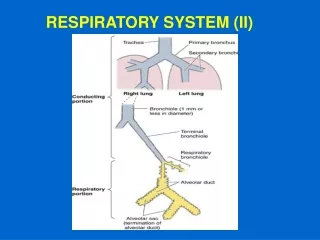
Anatomical division • upper respiratory tract • nasal cavity • paranasal cavities • nasopharynx • lower respiratory tract • larynx • trachea • tracheobronchial tree • respiratory compartment
Surgical division Anatomical division • upper respiratory tract • nasal cavity • paranasal cavities • nasopharynx • larynx • lower respiratory tract border: apertura thoracis sup. • trachea • tracheobronchial tree • respiratory compartment • upper respiratory tract • nasal cavity • paranasal cavities • nasopharynx • lower respiratory tract • larynx • trachea • tracheobronchial tree • respiratory compartment
General structure of respiratory system wall • tunica mucosa (mucosa) • epithelium • ciliated pseudostratified columnar (respiratory epithelium) • non-keratinized stratified squamous • lamina basalis • lamina propria • glands (seromucinous tuboalveolar), lymph nodes (noduli lymphoidei) • tunica fibromusculocartilaginea • collagenous and elastic tissue (and its ligaments – larynx, trachea) • smooth muscles (trachea, bronchi, bronchioli) • skeletal muscles (larynx) • tunica serosa or tunica adventitia • tunica serosa (pleura) has three layers: • mesothelium • lamina basalis • lamina propria • tela subserosa
Trachea pars cervicalis (C6- C7) pars thoracica (T1-T4) newborn at the level of C4, child C5 bifurcatio tracheae (T4) = 1st branching of tracheobronchial tree carina tracheae calibers: length 10-11 cm, width 25 mm syntopy: ventrally thyroid gland, dorsally oesophagus
Trachea – supply Arteries: a. thyroidea inf. → rr. tracheales aorta thoracica → rr. bronchiales (a. thyroidea ima – 2 %) newborns and children – branches from thymus arteries Veins: drain into oesophageal veins, into plexus thyroideus impar and into v. brachiocephalica sin. Lymph: nodi tracheobronchiales, nodi tracheales → truncus bronchomediastinalis dx.+ sin. Nerves: n. vagus → n. laryngeus recurrens truncus sympathicus
Coniotomy (coniopuncture) emergency procedure in outdoor (rare) transversal section between cartilago thyroidea et cricoidea through lig. cricothyroideum medianum Approach passes through following layers: skin + subcutaneous tissue lamina superficialis fasciae cervicalis lamina pretrachealis fasciae cervicalis lig. cricothyroideum medianum + mucosa !Cave! – interconnection of rr. cricothyroidei a. laryngeae superioris below cartilago thyroidea – lobus pyramidalis glandulae thyroideae (40%)
Tracheotomy sagittal section for canylation through several tracheal cartilages (done in hospital) tracheostomia superior above isthmus glandulae thyroideae (in the extent of cartilago trachealis 2-4) tracheostomia inferior below isthmus, above incisura jugularis Approachpasses through following layers: skin + subcutaneous tissue lamina superficialis fasciae cervicalis venous arcus venosus jugularis (only in lower tracheotomy) lamina pretrachealis fasciae cervicalis + cutting through midline fibrous connection of both mm. sternohyoidei (only in lower tracheotomy) venous plexus thyroideus impar (only in lower tracheotomy) cartilagines tracheales + ligg. anularia + mucosa
coniotomy • upper tracheotomy • lower tracheotomy
Tracheotomy – risks bleeding from: plexus thyroideus impar a. thyroidea ima (2%) arcus venosus jugularis lobus pyramidalis glandulae thyroideae (40%)
Trachea – wall structure epithelium of respiratory tract glandulae tracheales – seromucous glands cartilagines tracheales (15-20) C-shaped rings ligg. anularia / trachealia paries membranaceus – dorsal wall m. trachealis – smooth (horizontal as well as longitudinal fibers) adventicia on the surface
Tracheobronchial tree (Arbor bronchialis) 23 divisions –dichotomic branching primary bronchi (bronchus pricipalisdx.+ sin.) right: shorter, wider, straighter foreign body enters in 75% into the right one secondary bronchi (bronchi lobares) 2 left and 3 right tertiary bronchi (bronchi segmentales) 8 on the left and 10 on the right left: 1+2 connected, 7+8 connected in 90% exception: 6th bronchus segmentalis of both sides branches in trichotomic way!
Tracheobronchial tree (Arbor bronchialis) bronchi 4th order(bronchus subsegmentalis): b = ventral, a = dorsal bronchi 5th order: ii = ventral, i = dorzal bronchi 6th order: β = ventral, α = dorzal terminal bronchioli (bronchiolus terminalis) = 14th-16th order(originate by 14th division) 1 bronchiolus terminalis = 1 secondary pulmonary lobulus (visible on the lung surface) alveolar tree (originate by 17th branching)
Bronchi (Bronchi) tunica mucosa: epithelium of respiratory tract pseudostratified columnar with cilia seromucous glands tunica fibromusculocartilaginea: cartilages have irregular shape (more peripheraly disappear) smooth muscle – spiral (more peripheraly increases) nodes of lymphoid tissue – at the branching
Bronchioli (Bronchioli) caliber< 1 mm epithelium changes into simple cuboidal exocrinocyti bronchiolares (Clara cells) produce constituents of surfactant, lysosomal activity, mitoticactivity no cartilage, glands and lymph nodes increase of elastic fibers 1 bronchiolus terminalis = 1 secondary pulmonary lobule
Lungs (Pulmo) description: basis, apex facies costalis (+ pars vertebralis) facies mediastinalis (+ impressio cardiaca) facies diaphragmatica (facies interlobaris) margo anterior (incisura cardiaca p.sin.) margo inferior hilum pulmonis, radix pulmonis fissura obliqua, fissura horizontalis p. dx. impressions
Lungs – division hilum (clinically hilus) structures: left „ABV“ - right „BAV“ right lung – 3 lobes (sup., middle, inf.) left lung – 2 lobes (sup., inf.) lingula p. sin. segments (segmenta bronchopulmonalia) 10 on the right 10 on the left (sometimes 8) I+II fused, VII missing in 90%
HILUM PULMONIS Left lung Right lung A B V B A V
Lungs – impressions sulcus arteriae subclaviae (facies mediastinalis) impressio costae primae (margo anterior) impressiones costarum (facies costalis) impressio cardiaca (facies mediastinalis) Left lung: all on facies mediastinalis sulcus aorticus impressio oesophagea sulcus venae brachiocephalicae sinistrae Right lung: all on facies mediastinalis sulcus venae cavae superioris sulcus venae azygos (!correctly s.v. azygoi !) sulcus oesophageus
Blood supply of lungs – functional circuit right heart ventricle (deoxygenated blood)→ truncus pulmonalis → arteria pulmonalisdx.+ sin. → branching correspond to bronchi left hyparterial bronchus, right eparterial bronchus elastic arteries low-pressure vasculature 25/5 Torr smooth muscle cells in fetus, in adults since < 1 mm → capillaries (continuous) → oxygenated blood → venules independent on arteries in septa between lobules → 4 venae pulmonales (2 right and 2 left) → left heart atrium
Blood supply of lungs functional circuit arterio-venous anastomoses arterio-arterial anastomoses veno-venous anastomoses during hypoxia fastly growing arterial smooth muscle→ hypertrophy of right ventricle
Vascular supply of lungs – nutritive circuit aorta thoracica → rami bronchiales 1 right – usually from a. intercostalis tertia 2 left – directly from thoracic aorta → along bronchi as far as bronchioli respiratorii (rami bronchiales accessorii within lig. pulmonale) venae bronchiales deep system opening into vv. pulmonales superficial system drains blood from extrapulmonary bronchi, pleura and hilar lymphnodes → vv. pulmonales or v. azygos / hemiazygos accessoria
Lungs – lymph drainage superficial subpleural plexus deep plexus around bronchi and vessels Lung alveoli have no lymph vessels in their walls nodi lymphoidei intrapulmonales→ n.l. bronchopulmonales→ n.l. tracheobronchiales inferiores (both lungs except of three left upper segments I+II, III) → n.l. tracheobronchiales sup. dx. → truncus bronchomediastinalis dx. → angulus venosus dx. → v. brachiocephalica dx. I+II, III segments on the left – directly into n.l. tracheobronchiales sin. → truncus bronchomediastinalis sin. → ductus thoracicus → angulus venosus sin. → v. brachiocephalica sin.
Lungs – innervation nn. vagi both sides viscerosensory + autonomic parasympathetic stimuli truncus sympathicus autonomic sympathetic stimuli
Tracheobronchial tree (Arbor alveolaris) • dichotomic branching • from bronchioli respiratorii onwards • 17th-23rd order • functionally respiratory compartment
Tracheobronchial tree (Arbor alveolaris) respiratory bronchioles (bronchioli respiratorii) 17th-19th order (originating by 17th branching) pulmonary alveoli evaginate from their walls 19th order forms lobulus pulmonis primarius (8 primary lobuli together form one secondary) alveolar ducts (ductus alveolares) 20th-22nd order pulmonary alveoli evaginate from their walls at the end of 3rd orderalveolar duct there is atrium (atrium), divided by last, 23rd branching into two: alveolar saccules (sacculi alveolares) 23rd order evaginate only into: pulmonary alveoli (alveoli pulmonis)
Respiratory bronchioliBronchioli respiratorii diameter < 0,3 mm simple ciliated cuboidal epithelium branching of pulmonary alveoli continue into alveolar ducts
Lung alveoliAlveoli pulmonis 200 μm size, polyedric, thin-walled alveolar mucosa = respiratory epithelium alveolar septum alveolar pores (Kohn)
Alveolar mucosa pneumocytus typus I (pneumocyte type I, type I alveolar cell; membranous p.) 95% of mucosa flat, thin (25 nm) organels around nucleus pinocytic vesicles pneumocytus typus II(pneumocyte type II, type II alveolar cell; granular, spetal, great alveolar cells) ovoid shape with microvilli secretory structure (Mit, GER, GA) lamellar bodies (1,5 μm) = surfactant proliferate and differentiate (recovery of mucosa)
Interalveolar septumSeptum interalveolare cells fibroblasts (collagen type I and III) – septum cells endothelial cells of capillaries alveolar macrophages (macrophaygocyti alveolares) reticular and elastic fibers alveolar pores (pori septales) – 10 μm