Download
1 / 34
340 likes | 579 Vues
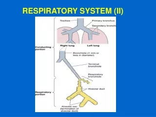
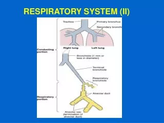
RESPIRATORY SYSTEM (II). EXTRAPULMONARY خارج الرئه BRONCHUS (primry BRONCHUS). Generally have the same histological appearance as the trachea. اذا راجع الدرس السابق للتفاصيل. INTRAPULMONARY داخل الرئه BRONCHI (2ry & 3ry BRONCHI). 1- Mucosa. 2- Muscle coat. 3- Submucosa.

E N D
EXTRAPULMONARY خارج الرئه BRONCHUS(primry BRONCHUS) Generally have the same histological appearance as the trachea. اذا راجع الدرس السابق للتفاصيل
INTRAPULMONARY داخل الرئه BRONCHI(2ry & 3ry BRONCHI) 1- Mucosa. 2- Muscle coat. 3- Submucosa. 4- Adventitia. تتكون هيستولوجيا من تفصل لاحقا
INTRAPULMONARY BRONCHUS • Mucosa: It has longitudinal طولي mucosal folds. a- Epithelium: Respiratory epith. b- L.P.: (Lamina propria) يتكون من Fibroelastic C.T. (loose C.T. rich in elastic fibers). It contains seromucous glands. “ “ lymphoid elements. N.B. No elastic lamina الدكتور يقول كلمه ( نو ) معناها سؤال يعني ركز عليها .
INTRAPULMONARY BRONCHUS (2) Muscle coat (complete): Two distinct مختلفتين layers of SMF ( smooth muscle fibers ) spirally حلزوني arranged in opposite direction (crisscrossing متقاطعه bundles of spirally arranged smooth muscle fibers “SMF”). (3) Submucosa: C.T. contains: a- Seromucous glands. b- Lymphoid elements.
INTRAPULMONARY BRONCHUS (4) Adventitia: Contents: a- Loose C.T.: Contains radially arranged elastic fibers to connect with counterparts of neighboring bronchial tree. b- Irregular plates of hyaline cartilage (complete layer). c- Solitary lymphoid nodules. اي شيء احمر وعليه خط هذا ركز عليه الدكتور وهذا في كل المحاضره
BRONCHIOLES لا يوجد بها غضاريف ولا بالتي بعدها عشان كذا من هنا انسى شيء اسمه غضاريف 1- Preterminal قبل النهائيه( 1ry ) Bronchioles (Bronchioles): Are less than 1mm in diameter. Each bronchiole supplies pulmonary lobule. 2- Terminal النهائيه ( 2ry ) Bronchioles. 3- Respiratory ( 3ry ) Bronchioles.
Preterminal Bronchioles (1) Mucosa: has longitudinal folds: A- Epithelium: 1- Simple ciliated columnar epith مهمه جدا معرفه نوع الابيثيلويم لانه محل مقارنه لانه سوف يتغير في التراكيب القادمه. with occasional goblet cells (in larger preter. br.). 2- مهمه: تركيب الجزء الاخير منها Simple cuboidal mostly ciliated with occasional Clara cells BUT NO goblet cells(in smaller preter. bronchioles). B- Lamina propria: FibroelasticC.T. (rich in elastic Fs.) (2) Smooth muscle: 2 helically arranged SM layers. (3) Adventitia: loose fibroelastic C.T. N.B مهمه جدا جدا. No cartilage, No seromucous glands, No lymphnoduLes.
Terminal Bronchioles Similar structure to preterminal. bronchioles, but: Epithelium: Simple cuboidal partially ciliated epithelium With Clara cells. مثل تركيب الجزء الاخير من السابقه N.B. Are less than 0.5mm in diameter. N.B. Each supplies lung acinus.
Respiratory Bronchioles Are similar in structure to terminal bronchiolesBut: their walls are interrupted by the presence of few pulmonary alveoli. مهمه جدا وخصوصا بالعملي
Clara cells خلايا مرطبه تقوم مقام القلوبليت
CLARA CELLS Structure: columnar cells (non ciliated). Dome-shaped apices with microvilli. Numerous apical secretory granules (of glycoproteins). Abundant rER. Function: مهمه 1- Protect the bronchiolar epith. by their glycoproteins secretion. 2- Degrade toxins in inhaled air. 3- Divide to regenerate the bronchiolar epith. 4- Produce surfactant-like material. مواقع انتشارها ( مهمه جدا ) : 1- last portion of preterminal bronchioles 2- all terminal bronchioles 3- all respiratory portion
ALVEOLAR DUCTS The wall of alveolar ducts consist almost of pulmonary alveoli. Alveolar ducts do NOT have walls of their own; They are merely فقط linear arrangement of pulmonary alveoli. N.B. Alveolar duct → ends by: atrium → communicates with: 2-3 alveolar sacs
PULMONARY ALVEOLI Definition: They are small outpouchings تجيّب خارجي of respiratory bronchioles, alveolar ducts & alveolar sacs. Topics: العنوانين التي ستتم مناقشتها *Interalveolar septa. *Blood-air barrier ( Blood-gas barrier) *Alveolar epithelium. *Lung macrophages (alveolar macrophages).
1- INTERALVEOLAR SEPTA Definition: The region between 2 adjacent متجاوه alveoli. Components: • Alveolar Epithelium: lines both sides of interalveolar septum. (B) Interstitium.
ALVEOLAR EPITHELIUM • Type I Pneumocytes (Type I alveolar cells) (Squamous alveolar cells). (2) Type II Pneumocytes (Type II alveolar cells) ( Septal cells) ( Great alveolar cells)
ALVEOLAR EPITHELIUM Type I Pn. Type II Pn.
ALVEOLAR EPITHELIUM • Type I Pneumocytes: - line 95% of the alveolar surface. - Count:less numerous لان حجمها كبير than type II pneumocytes. - L/M: simple squamous epith. ,highly attenuated cells. - E/M: Abundant pinocytotic vesicles, Are connected together and with type II cells by occluding junctions. -Function: Exchange of gases.
(2) Type II Pneumocytes: • Line 5% of the alveolar surfaces. • Are more numerous than type I pneumocytes. • L/M: Are cuboidal cells ( other textbooks: rounded cells).Usually found in groups of 2-3 cells. Usually found at sites of union of septa. Foamy منتفخ- رائغ or vesicular cytoplasm. Nucleus: central, rounded, vesicular. هذا النوع من الخلايا هو المسؤول عن التعويض فهو يعوض خلايا تايب 1 وتايب 2
Type II Pneumocytes: • E/M: connected with type I cells by occluding junctions Dome-shaped قبي الشكل apical surface. Short apical microvilli. Abundant mitochondria, RER , Well-developed Golgi. Membrane-bound Lamellar bodies (contain concentric or parallel lamellae limited by a unit membrane) (contain pulmonary surfactant).
Type II Pneumocytes: Function: 1- Synthesis & secretion of pulmonary surfactant التي وظائفه : a- It reduces effort to inflate pulm. Alveoli. b- It has bactericidal effect. 2- Phagocytosis of pulmonary surfactant. 3-Renewal of alveolar epithelial cells: Type II cells can divide to regenerate both type I & type II pneumocytes.
Interstitium of interalveolar septa • Continuous Pulmonary Capillaries: -The richest capillary network in the body - Continuous blood capillaries - Endothelium shows numerous pinocytotic vesicles. (2) Interstitial C.T.: a- C.T. Fibers: elastic fibers & type III collagen (reticular fibers). b- C.T. Cells: Fibroblasts, Macrophages, Mast cells, Lymphocytes.
BLOOD-GAS BARRIER(BLOOD-AIR BARRIER) Definition: It is the region of the interalveolar septum that is traversed by O2 & CO2 Components: 1- Thin layer of surfactant. 2- Type I pneumocyte. 3- Fused basal laminae of type I pneumocytes & endothelial cells of the pulmonary capillary. 4- Endothelial cells of the pulmonary capillary.
Alveolar Macrophages(Dust Cells) Sites: • In lumen of pulmonary alveoli. • In pulmonary interstitium. Function: 1- Phagocytose particulate matter (e.g. dust & bacteria)in the lumen of pulm. alveoli & in the interalveolar septa. 2- Phagocytose part of the surfactant.