Chapter 13 Respiratory System
Chapter 13 Respiratory System. Anatomy and Physiology II Ms . Harborth. Organs of the Respiratory System. Nose Pharynx Larynx Trachea Bronchi Lungs – alveoli. Function of the Respiratory System. Oversees gas exchanges between the blood and external environment

Chapter 13 Respiratory System
E N D
Presentation Transcript
Chapter 13Respiratory System Anatomy and Physiology II Ms. Harborth
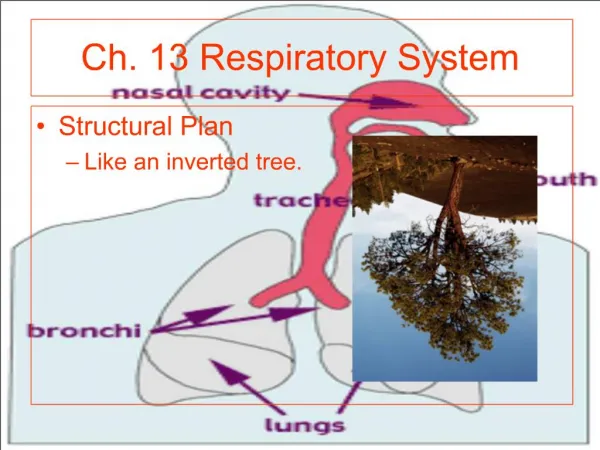
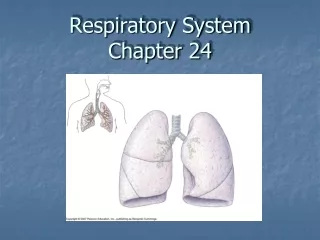
Organs of the Respiratory System Nose Pharynx Larynx Trachea Bronchi Lungs – alveoli
Function of the Respiratory System Oversees gas exchanges between the blood and external environment Exchange of gasses takes place within the lungs in the alveoli Passageways to the lungs purify, warm, and humidify the incoming air
The Nose The only externally visible part of the respiratory system Air enters the nose through the external nares (nostrils) The interior of the nose consists of a nasal cavity divided by a nasal septum
Anatomy of the Nasal Cavity • Olfactory receptors are located in the mucosa on the superior surface • The rest of the cavity is lined with respiratory mucosa • Lateral walls have projections called conchae • The nasal cavity is separated from the oral cavity by the palate • Anterior hard palate (bone) • Posterior soft palate (muscle)
Paranasal Sinuses • Function of the sinuses • Lighten the skull • Act as resonance chambers for speech • Produce mucus that drains into the nasal cavity • Cavities within bones surrounding the nasal cavity
Checkpoint! What are the nostrils referred to as? What separates the nostrils? Describe the two palates. Name one function of the paranasal sinuses.
Pharynx (Throat) • Muscular passage from nasal cavity to larynx • Three regions of the pharynx • Nasopharynx – superior region behind nasal cavity • Oropharynx – middle region behind mouth • Laryngopharynx – inferior region attached to larynx • The oropharynx and laryngopharynx are common passageways for air and food
Structure of the Pharynx • Auditory tubes enter the nasopharynx • Tonsils of the pharynx • Pharyngeal tonsil (adenoids) in the nasopharynx • Palatine tonsils in the oropharynx • Lingual tonsils at the base of the tongue
Larynx (Voice Box) Routes air and food into proper channels Plays a role in speech Made of eight rigid hyaline cartilages and a spoon-shaped flap of elastic cartilage (epiglottis)
Structures of the Larynx • Thyroid cartilage • Largest hyaline cartilage • Protrudes anteriorly (Adam’s apple) • Epiglottis • Superior opening of the larynx • Routes food to the larynx and air toward the trachea • Vocal cords (vocal folds) • Vibrate with expelled air to create sound (speech) • Glottis – opening between vocal cords
Trachea (Windpipe) Connects larynx with bronchi Lined with ciliated mucosa Walls are reinforced with C-shaped hyaline cartilage
Checkpoint! What are the three regions of the pharynx? What routes food into the esophagus? What cartilage structure forms the “Adam’s apple?” Explain the function of the ciliated cells lining the trachea.
Primary Bronchi Formed by division of the trachea Enters the lung at the hilus(medial depression) Right bronchus is wider, shorter, and straighter than left Bronchi subdivide into smaller and smaller branches
Lungs • Occupy most of the thoracic cavity • Apex is near the clavicle (superior portion) • Base rests on the diaphragm (inferior portion) • Each lung is divided into lobes by fissures • Left lung – two lobes • Right lung – three lobes
Coverings of the Lungs Pulmonary (visceral) pleura covers the lung surface Parietal pleura lines the walls of the thoracic cavity Pleural fluid fills the area between layers of pleura to allow gliding
Bronchioles Smallest of bronchi Terminal bronchioles end in alveoli
Respiratory Zone • Structures • Respiratory bronchioli • Alveolar duct • Alveoli • Site of gas exchange
Alveoli • Structure of alveoli • Alveolar duct • Alveolar sac • Alveolus • Gas exchange takes place within the alveoli in the respiratory membrane
Gas Exchange • Gas crosses the respiratory membrane by diffusion • Oxygen enters the blood • Carbon dioxide enters the alveoli • Macrophages add protection • Surfactant coats gas-exposed alveolar surfaces
When Things Go Wrong Sinusitis Tonsillitis Smoker’s cough Choking Cleft palate Pleurisy
Checkpoint! What does the trachea branch into? What do the bronchi branch into? Where does gas exchange take place? What is pleurisy?
Respiration Events ALL FOUR ARE NECESSARY FOR GAS EXCHANGE Pulmonary ventilation External respiration Respiratory gas transport Internal respiration
Mechanics of Breathing (Pulmonary Ventilation) • Depends on volume change in thoracic cavity • Two phases • Inspiration – flow of air into lung • Diaphragm and intercostal muscles contract • The size of the thoracic cavity increases, air is pulled into lungs • Expiration – air leaving lung • Largely a passive process which depends on natural lung elasticity • As muscles relax, air is pushed out of the lungs • Forced expiration can occur mostly by contracting internal intercostal muscles to depress the rib cage
Nonrespiratory Air Movements • Can be caused by reflexes or voluntary actions • Examples • Cough and sneeze – clears lungs of debris • Laughing • Crying • Yawn • Hiccup
Respiratory Volumes and Capacities • Normal breathing moves about 500 ml of air with each breath (tidal volume [TV]) • Many factors that affect respiratory capacity • A person’s size, sex, age, physical condition • Residual volume of air – after exhalation, about 1200 ml of air remains in the lungs
Respiratory Volumes and Capacities • Inspiratory reserve volume (IRV) • Amount of air that can be taken in forcibly over the tidal volume • Usually between 2100 and 3200 ml • Expiratory reserve volume (ERV) • Amount of air that can be forcibly exhaled • Approximately 1200 ml • Vital capacity • The total amount of exchangeable air • Vital capacity = TV + IRV + ERV • Functional volume • Air that actually reaches the respiratory zone • Usually about 350 ml (150 ml = dead space volume) • Respiratory capacities are measured with a spirometer
Respiration Sounds • Sounds are monitored with a stethoscope • Bronchial sounds – produced by air rushing through trachea and bronchi • bronchial sounds • Vesicular breathing sounds – soft sounds of air filling alveoli • vesicular sounds
Checkpoint! Name one of the events that must take place for respiration to occur. What happens when your diapraghm and external intercostals contract? What happens when they relax? What is IRV? What is TV?
External Respiration • O2movement into the blood • The alveoli always has more O2 than the blood • O2moves by diffusion towards the area of lower concentration • Pulmonary capillary blood gains O2 • CO2 movement out of the blood • Blood returning from tissues has higher concentrations of CO2 than air in the alveoli • Pulmonary capillary blood gives up CO2 • Blood leaving the lungs is O2 -rich and CO2 -poor
Gas Transport in Blood • Oxygen transport in the blood • Inside red blood cells attached to hemoglobin (oxyhemoglobin [HbO2]) • A small amount is carried dissolved in the plasma • Carbon dioxide transport in the blood • Most is transported in the plasma as bicarbonate ion (HCO3–) • Small amount is carried on hemoglobin, but at different binding sites than those of oxygen
Internal Respiration • Exchange of gases between blood and body cells • An opposite reaction to what occurs in the lungs • Carbon dioxide diffuses out of tissue to blood • Oxygen diffuses from blood into tissue
Neural Regulation of Respiration Activity of respiratory muscles is transmitted to the brain by the phrenic and intercostal nerves Neural centers that control rate and depth are located in the medulla The pons appears to smooth out respiratory rate Normal respiratory rate (eupnea) is 12–15 respirations per minute Hyperniais increased respiratory rate often due to extra oxygen needs
Factors Influencing Respiratory Rate and Depth • Physical factors • Increased body temperature • Exercise • Talking • Coughing • Volition (conscious control) • Emotional factors • Chemical factors • Carbon dioxide levels • Main regulatory chemical for respiration • Increased carbon dioxide increases respiration • Changes in carbon dioxide act directly on the medulla oblongata • Oxygen levels • Changes in oxygen concentration in the blood are detected by chemoreceptors in the aorta and carotid artery • Information is sent to the medulla oblongata
COPD • Chronic Obstructive Pulmonary Disorder • Chronic Bronchitis and Emphysema • Patients almost always have a history of smoking • Labored breathing (dyspnea) becomes progressively more severe • Coughing and frequent pulmonary infections are common • Ultimately develop respiratory failure
Emphysema Alveoli enlarge as adjacent chambers break through Chronic inflammation promotes lung fibrosis Airways collapse during expiration Patients use a large amount of energy to exhale Overinflationof the lungs leads to a permanently expanded barrel chest Cyanosis
Lung Cancer • Accounts for 1/3 of all cancer deaths in the United States • Increased incidence associated with smoking • Three common types • 1. Squamous cell carcinoma • 2. Adenocarcinoma • 3. Small cell carcinoma
SIDS • Sudden Infant Death Syndrome • Apparently healthy infant stops breathing and dies during sleep • Some cases are thought to be a problem of the neural respiratory control center • One third of cases appear to be due to heart rhythm abnormalities
Asthma asthmatic wheeze Chronic inflamed hypersensitive bronchiole passages Response to irritants with dyspnea, coughing, and wheezing
Developmental Aspects • Lungs are filled with fluid in the fetus • Lungs are not fully inflated with air until two weeks after birth • Surfactant that lowers alveolar surface tension is not present until late in fetal development and may not be present in premature babies • Important birth defects • Cystic fibrosis – oversecretion of thick mucus clogs the respiratory system • Cleft palate