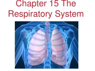
Respiratory System Chapter 15
Respiratory System Chapter 15. Created by; Nicole Manago and Morgan Baldy. Respiratory System. Respiratory System. Respiratory System Functions. Nose- Where air normally enters the Respiratory system.

Respiratory System Chapter 15
E N D
Presentation Transcript
Respiratory SystemChapter 15 Created by; Nicole Manago and Morgan Baldy
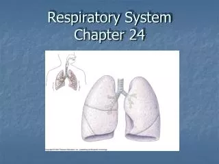
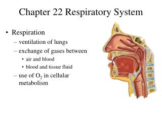
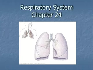
Respiratory System Functions • Nose- Where air normally enters the Respiratory system. • Pharynx- The throat, extends between the internal nares and entrance to larynx / esophagus. • Larynx- Inhaled air leaves the pharynx and enters larynx (voice box). • Trachea- Helps contraction to make large volumes of air along passageways. • Bronchi/ Bronchioles- Supplies the lungs with air. • Alveoli- Gives the lungs an open spongy appearance. • Lungs- How one breathes, the right lung has 3 lobes and the left lung has 2 lobes. • Pleural Cavity- Rib cage and muscular diaphragm, each lung occupies a cavity.
Pulmonary Ventilation: Pressure Changes • Pulmonary Ventilation is the physical movement of air into and out of the respiratory tract. • The volume of the thoracic cavity increases when the ribs are elevated and the diaphragm is depressed during contraction. • When the rib cage and diaphragm are at rest the pressures inside and outside are equal and no air movement occurs.
Pulmonary Ventilation: Pressure Changes • During inhalation, elevation of the rib cage and depression of the diaphragm increases the volume of the thoracic cavity. Pressure in the lungs decrease and air flows into the lungs. • During exhalation, the rib cage returns to its original position or the diaphragm relaxes. Reducing the volume of the thoracic cavity, pressure in the lungs rises and air flows out of the lungs. • During both inhalation and exhalation contraction of accessory muscles may assist movements of the rib cage to increase depth and rate of respiration.
Lung Volumes • The amount of air moved into or out of the lungs during a single respiratory cycle is the tidal volume. • Expiratory Reserve Volume - The amount of air that can be voluntarily expelled at the end of a respiratory cycle • Inspiratory reserve volume(IRV) - The amount of air that can be taken in over and above the resting tidal volume. Since male and female lung capacities are much different, a males IRV will average 3300 mL while a female is 1900mL.
Lung Volumes • Vital Capacity - The sum of the inspiratory reserve volume, the expiratory reserve volume, and the total volume. • It is also the maximum amount of air that can be moved in and out of the lungs in one respiratory cycle. • Residual Volume - The amount of air that remains in your lungs even after a maximal exhalation. • A reason why this exists is because the lungs are held against the thoracic wall and this prevents the elastic fibers from contracting any further. • Minimal Volume - When a chest cavity has been penetrated, the lungs will collapse and the amount of air is reduced to the minimal volume.
Oxygen Transport • Oxygen is transported bound to Hemoglobin Molecules • Specifically to the iron ions in the center of heme units • This process occurs through a reversible reaction • Hb+O2 HbO2 • The amount of oxygen released by hemoglobin depends primarily on the Po2 of its surroundings • The lower the oxygen content of a tissue, the more oxygen is released by hemoglobin molecules passing through local capillaries • Active tissues will receive roughly three times much oxygen as inactive tissues • The amount of oxygen released is also influenced by pH and temperature
Oxygen Transport • Active Tissues generate acids that lower the pH of the interstitial fluids • When the pH declines, hemoglobin molecules release their bound oxygen molecules faster • Po2, pH, and temperature re important during periods of maximal exertion • The combination of the three factor-s makes the hemoglobin entering the area release much more oxygen that can release much more oxygen that can be used by active muscle fibers
Carbon Dioxide Transport • Carbon Dioxide is generated by aerobic metabolism in peripheral tissues • After entering the bloodstream, a CO2 Molecule may: • Dissolve in Plasma • Bind to hemoglobin within red blood cells • Be converted to a molecule of carbon acid (H2CO3) • Only about 7% of the carbon dioxide absorbed by the peripheral capillaries is transported as dissolved gas molecules • In the red blood cells, some of the carbon dioxide molecules are bound to the protein “globin” portions of the hemoglobin molecules • This forms carbaminohemoglobin • 23% of the carbon dioxide entering the blood in the peripheral tissues is transported as carbaminohemoglobin
Carbon Dioxide Transport • 70% of all carbon dioxide molecules in the body are transported in the plasma as bicarbonate ions • Carbon Dioxide in Red Blood Cells is converted to carbonic acid by the enzyme carbonic anhydrase • Each molecule dissociates into a hydrogen ion and a bicarbonate ion • Co2+H2O H2CO3 H+ + HCO3- • Hydrogen Ions bind to hemoglobin molecules, preventing both their release from the RBCs and lowering of plasma pH • Bicarbonate ions diffuse into surrounding plasma • When venous blood reaches the alveoli, carbon dioxide diffuses out of plasma, and P CO2 declines. • The transport of oxygen and carbon dioxide in the blood involves reactions that are completely reversible
Respiratory Control • If the peripheral tissue becomes more active, the interstitial Po2 falls and the P co2 rises • This makes for more oxygen being delivered and more carbon dioxide being carried away • Rising Pco2 levels case the relaxation of smooth muscles in the walls of arterioles in the area, increasing blood flow • When Pco2 increases, the bronchioles dilate; when the Pco2 declines the bronchioles constrict • Airflow is directed to lobules in which the Pco2 is high
Respiratory Control • Both voluntary and involuntary • The brains involuntary respiratory centers regulates the respiratory muscles and control the frequency and the depth of breathing • The voluntary control of respiration reflects activity in the cerebral cortex that affects the out of the respiratory centers or of motor neurons that control respiratory muscles • Respiratory Centers- are three pairs of nuclei in the reticular formation of the pons and medulla oblongata • The respiratory rhythmicity centers set the pace for respiration
Reflex Control • Breathing occurs without conscious control • Activities of the respiratory centers are modified by sensory information from mechanoreceptors and chemoreceptors • Mechanoreceptors respond to changes in lung volume or to changes in arterial blood pressure • The inflation reflex- prevents the lungs from over expanding during forced breathing • The mechanoreceptors involved are stretch receptors that are stimulated when the lungs expand • As volume of the lungs increase, the DRG inspiratory center is gradually inhibited, and the VRG expiratory center is stimulated • Deflation reflex- inhibits the expiratory center and stimulates inspiratory center when the lungs are collapsing • Output from these baroreceptors also affects the respiratory centers • When blood pressure falls, the respiratory rate increases: when blood pressure rises, the respiratory rate declines
Chemoreceptor Reflexes • They respond to chemical changes in the blood and cerebrospinal fluid. • The stimulation leads to an increase in the depth and rate of respiration • Receptors in the medulla oblongata respond to the pH and Pco2 in cerebrospinal fluid • Carbon Dioxide levels have a more powerful effect on respiratory activity than do oxygen levels • This is because a relatively small increase in arterial Pco2 stimulates CO2 receptors, but arterial Po2 doesn't’t usually decline enough to activate oxygen receptors • Carbon Dioxide levels regulate respiratory activity under normal conditions • The cooperation between the carbon dioxide and oxygen receptors breaks down only under unusual circumstances • If Po2 is reduced enough, breath-holding ability may increase to the point that the individual becomes unconscious from oxygen starvation in the brain without ever feeling urge to breath • The chemoreceptors monitoring CO2 levels are also sensitive to pH • Any condition affecting pH of blood or CSF will affect respiratory performance