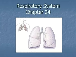
Chapter 23 Respiratory System
Chapter 23 Respiratory System. Albert Grazia, M.S., N.D. (516) 486-8332 www.naturedoc.info. Chapter 23 The Respiratory System. Cells continually use O2 & release CO2 Respiratory system designed for gas exchange Cardiovascular system transports gases in blood Failure of either system


Chapter 23 Respiratory System
E N D
Presentation Transcript
Chapter 23 Respiratory System Albert Grazia, M.S., N.D. (516) 486-8332 www.naturedoc.info Albert Grazia, M.S., N.D. www.naturedoc.info
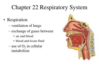
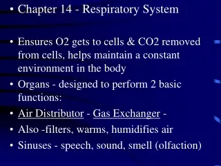
Chapter 23The Respiratory System • Cells continually use O2 & release CO2 • Respiratory system designed for gas exchange • Cardiovascular system transports gases in blood • Failure of either system • rapid cell death from O2 starvation
Respiratory System Anatomy • Nose • Pharynx = throat • Larynx = voicebox • Trachea = windpipe • Bronchi = airways • Lungs • Locations of infections • upper respiratory tract is above vocal cords • lower respiratory tract is below vocal cords
STRUCTURE FUNCTION nose / nasal cavity nose/ nasal cavity warms, moistens, & filters air as it is inhaled pharynx (throat) pharynx (throat) passageway for air, leads to trachea passageway for air, leads to trachea larynx larynx the voice box, where vocal chords are located the voice box, where vocal chords are located Albert Grazia, M.S., N.D. www.naturedoc.info
trachea (windpipe) tube from pharynx to bronchi rings of cartilage provide structure, keeps the windpipe "open" trachea is lined with fine hairs called cilia which filter air before it reaches the lungs Albert Grazia, M.S., N.D. www.naturedoc.info
bronchi two branches at the end of the trachea, each lead to a lung bronchioles a network of smaller branches leading from the bronchi into the lung tissue & ultimately to air sacs alveoli the functional respiratory units in the lung where gases (oxygen & carbon dioxide) are exchanged (enter & exit the blood stream) Albert Grazia, M.S., N.D. www.naturedoc.info
External Nasal Structures • Skin, nasal bones, & cartilage lined with mucous membrane • Openings called external nares or nostrils
Nose -- Internal Structures • Large chamber within the skull • Roof is made up of ethmoid and floor is hard palate • Internal nares (choanae) are openings to pharynx • Nasal septum is composed of bone & cartilage • Bony swelling or conchae on lateral walls
Functions of the Nasal Structures • Olfactory epithelium for sense of smell • Pseudostratified ciliated columnar with goblet cells lines nasal cavity • warms air due to high vascularity • mucous moistens air & traps dust • cilia move mucous towards pharynx • Paranasal sinuses open into nasal cavity • found in ethmoid, sphenoid, frontal & maxillary • lighten skull & resonate voice
Rhinoplasty • Commonly called a “nose job” • Surgical procedure done for cosmetic reasons / fracture or septal repair • Procedure • local and general anesthetic • nasal cartilage is reshaped through nostrils • bones fractured and repositioned • internal packing & splint while healing Albert Grazia, M.S., N.D. www.naturedoc.info
Pharynx • Muscular tube (5 inch long) hanging from skull • skeletal muscle & mucous membrane • Extends from internal nares to cricoid cartilage • Functions • passageway for food and air • resonating chamber for speech production • tonsil (lymphatic tissue) in the walls protects entryway into body • Distinct regions -- nasopharynx, oropharynx and laryngopharynx
Nasopharynx From choanae to soft palate openings of auditory (Eustachian) tubes from middle ear cavity adenoids or pharyngeal tonsil in roof Passageway for air only pseudostratified ciliated columnar epithelium with goblet
Oropharynx From soft palate to epiglottis fauces is opening from mouth into oropharynx palatine tonsils found in side walls, lingual tonsil in tongue Common passageway for food & air stratified squamous epithelium
Laryngopharynx Extends from epiglottis to cricoid cartilage Common passageway for food & air & ends as esophagus inferiorly stratified squamous epithelium
Cartilages of the Larynx • Thyroid cartilage forms Adam’s apple • Epiglottis---leaf-shaped piece of elastic cartilage • during swallowing, larynx moves upward • epiglottis bends to cover glottis • Cricoid cartilage---ring of cartilage attached to top of trachea • Pair of arytenoid cartilages sit upon cricoid • many muscles responsible for their movement • partially buried in vocal folds (true vocal cords)
Larynx • Cartilage & connective tissue tube • Anterior to C4 to C6 • Constructed of 3 single & 3 paired cartilages
Vocal Cords • False vocal cords (ventricular folds) found above vocal folds (true vocal cords) • True vocal cords attach to arytenoid cartilages
The Structures of Voice Production • True vocal cord contains both skeletal muscle and an elastic ligament (vocal ligament) • When 10 intrinsic muscles of the larynx contract, move cartilages & stretch vocal cord tight • When air is pushed past tight ligament, sound is produced (the longer & thicker vocal cord in male produces a lower pitch of sound) • The tighter the ligament, the higher the pitch • To increase volume of sound, push air harder
Movement of Vocal Cords • Opening and closing of the vocal folds occurs during breathing and speech
Speech and Whispering • Speech is modified sound made by the larynx. • Speech requires pharynx, mouth, nasal cavity & sinuses to resonate that sound • Tongue & lips form words • Pitch is controlled by tension on vocal folds • pulled tight produces higher pitch • male vocal folds are thicker & longer so vibrate more slowly producing a lower pitch • Whispering is forcing air through almost closed rima glottidis -- oral cavity alone forms speech
Trachea • Size is 5 in long & 1in diameter • Extends from larynx to T5 anterior to the esophagus and then splits into bronchi • Layers: • mucosa = pseudostratified columnar with cilia & goblet • submucosa = loose connective tissue & seromucous glands • hyaline cartilage = 16 to 20 incomplete rings • open side facing esophagus contains trachealis m. (smooth) • internal ridge on last ring called carina • adventitia binds it to other organs
Trachea and Bronchial Tree • Full extent of airways is visible starting at the larynx and trachea
Histology of the Trachea • Ciliated pseudostratified columnar epithelium • Hyaline cartilage as C-shaped structure closed by trachealis muscle
Airway Epithelium • Ciliated pseudostratified columnar epithelium with goblet cells produce a moving mass of mucus.
Tracheostomy and Intubation • Reestablishing airflow past an airway obstruction • crushing injury to larynx or chest • swelling that closes airway • vomit or foreign object • Tracheostomy is incision in trachea below cricoid cartilage if larynx is obstructed • Intubation is passing a tube from mouth or nose through larynx and trachea Albert Grazia, M.S., N.D. www.naturedoc.info
Bronchi and Bronchioles • Primary bronchi supply each lung • Secondary bronchi supply each lobe of the lungs (3 right + 2 left) • Tertiary bronchi supply each bronchopulmonary segment • Repeated branchings called bronchioles form a bronchial tree
Histology of Bronchial Tree • Epithelium changes from pseudostratified ciliated columnar to nonciliated simple cuboidal as pass deeper into lungs • Incomplete rings of cartilage replaced by rings of smooth muscle & then connective tissue • sympathetic NS & adrenal gland release epinephrine that relaxes smooth muscle & dilates airways • asthma attack or allergic reactions constrict distal bronchiole smooth muscle • nebulization therapy = inhale mist with chemicals that relax muscle & reduce thickness of mucus
Pleural Membranes & Pleural Cavity • Visceral pleura covers lungs --- parietal pleura lines ribcage & covers upper surface of diaphragm • Pleural cavity is potential space between ribs & lungs
Gross Anatomy of Lungs • Base, apex (cupula), costal surface, cardiac notch • Oblique & horizontal fissure in right lung results in 3 lobes • Oblique fissure only in left lung produces 2 lobes
Mediastinal Surface of Lungs • Blood vessels & airways enter lungs at hilus • Forms root of lungs • Covered with pleura (parietal becomes visceral)
Structures within a Lobule of Lung • Branchings of single arteriole, venule & bronchiole are wrapped by elastic CT • Respiratory bronchiole • simple squamous • Alveolar ducts surrounded by alveolar sacs & alveoli • sac is 2 or more alveoli sharing a common opening
Histology of Lung Tissue Photomicrograph of lung tissue showing bronchioles, alveoli and alveolar ducts.
Cells Types of the Alveoli • Type I alveolar cells • simple squamous cells where gas exchange occurs • Type II alveolar cells (septal cells) • free surface has microvilli • secrete alveolar fluid containing surfactant • Alveolar dust cells • wandering macrophages remove debris
Alveolar-Capillary Membrane • Respiratory membrane = 1/2 micron thick • Exchange of gas from alveoli to blood • 4 Layers of membrane to cross • alveolar epithelial wall of type I cells • alveolar epithelial basement membrane • capillary basement membrane • endothelial cells of capillary • Vast surface area = handball court
Details of Respiratory Membrane • 4 layers comprise the respiratory membrane
Double Blood Supply to the Lungs • Deoxygenated blood arrives through pulmonary trunk from the right ventricle • Bronchial arteries branch off of the aorta to supply oxygenated blood to lung tissue • Venous drainage returns all blood to heart • Less pressure in venous system • Pulmonary blood vessels constrict in response to low O2 levels so as not to pick up CO2 on their way through the lungs
Breathing or Pulmonary Ventilation • Air moves into lungs when pressure inside lungs is less than atmospheric pressure • How is this accomplished? • Air moves out of the lungs when pressure inside lungs is greater than atmospheric pressure • Atmospheric pressure = 1 atm or 760mm Hg
There are no muscles in your lungs. They do not actively pump air in & out, in & out. The muscle responsible for breathing actually lies below the lungs. It is like a rubber sheet that separates your chest cavity & your abdominal cavity. It's name is diaphragm. • When you inhale, the diaphragm contracts & moves downward, which creates more space in your chest cavity & draws air into the lungs. When you exhale, the diaphragm relaxes & moves upward, forcing air out of the lungs. • A common demonstration of the mechanics behind breathing involves a bell jar, some glass tubing, and a couple of balloons. Like so: Albert Grazia, M.S., N.D. www.naturedoc.info
Mechanics of Breathing • To take a breath in, the external intercostal muscles contract, moving the ribcage up and out. The diaphragm moves down at the same time, creating negative pressure within the thorax. The lungs are held to the thoracic wall by the pleural membranes, and so expand outwards as well. This creates negative pressure within the lungs, and so air rushes in through the upper and lower airways. • Expiration is mainly due to the natural elasticity of the lungs, which tend to collapse if they are not held against the thoracic wall. This is the mechanism behind lung collapse if there is air in the pleural space (pneumothorax). Albert Grazia, M.S., N.D. www.naturedoc.info
Boyle’s Law • As the size of closed container decreases, pressure inside is increased • The molecules have less wall area to strike so the pressure on each inch of area increases.
Dimensions of the Chest Cavity • Breathing in requires muscular activity & chest size changes • Contraction of the diaphragm flattens the dome and increases the vertical dimension of the chest
Quiet Inspiration • Diaphragm moves 1 cm & ribs lifted by muscles • Intrathoracic pressure falls and 2-3 liters inhaled
Quiet Expiration • Passive process with no muscle action • Elastic recoil & surface tension in alveoli pulls inward • Alveolar pressure increases & air is pushed out
Labored Breathing • Forced expiration • abdominal mm force diaphragm up • internal intercostals depress ribs • Forced inspiration • sternocleidomastoid, scalenes & pectoralis minor lift chest upwards as you gasp for air