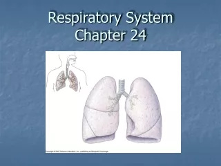
Overview of the Respiratory System: Key Components and Lung Structure
This chapter provides an in-depth overview of the respiratory system, detailing its primary components, including the nose, pharynx, larynx, trachea, bronchi, and lungs. It examines the structure of the trachea and main bronchi, highlighting their three essential layers: mucosa, submucosa, and adventitia. The chapter also describes lung anatomy, focusing on airways branching from the bronchi into bronchioles and alveoli. Key functions such as gas exchange and the role of various lung cells are explained, providing a clear understanding of respiratory physiology.

Overview of the Respiratory System: Key Components and Lung Structure
E N D
Presentation Transcript
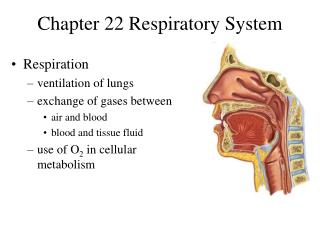
1. Components ---nose ---pharynx ---larynx ---trachea ---bronchi ---lung
2. Trachea and main bronchi three layers 1)Mucosa: ---epithelium: pseudostratified ciliated columnar epithelium ---lamina propria: CT, contain LC, PC, MC, BV, LV
Pseudostratified ciliated columnar epi. • ciliated cell: columnar, cilia • goblet cell • basal cell: -pyramidal, basally-located -undifferentiated cell→ciliated cell or goblet cell
brush cell: -columnar, microvilli, -EM: RER, no g. -function: not very clear, may be i.become into ciliated cell ii.receive sensory stimuli-epitheliodendritic synapse
diffuse neuroendocrine cell: -less, pyramidal -EM: dense-core g.-small granule cell neuroepithelial body: cell + NF -Function: secret hormones to regulate contract of SM and secretion of gland i. 5-hydroxytryptamine(serotonin) ii. calcitonin iii. enkephalin * clear basement membrane
2) Submucosa: LCT, with BV, LV and N • tracheal gland: mixed • diffuse LT and LN * S Ig A = secretory component (secreted by epi. cell) + Ig A ( produced by plasma cell)
3) Adventitia: • cartilage ring: 16-20 “C ” shaped • circular ligament: elastic F • SM- posterior part( membrane part): SM, elastic F, tracheal gland
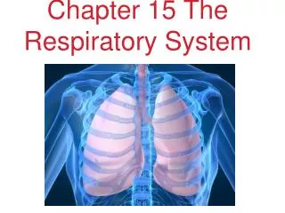
3. Lung ---paired organ, located in thoracic cavity
1) General structure: ---capsule: visceral layer of pleura- serous membrane-CT + mesothelium ---parenchyma: all branches of bronchi and alveoli( right 3, left 2) ---interstitia: CT, BV, LV, N
* branchi →intrapulmonary bronchial tree( lobar bronchial tree, segmental bronchi and small bronchi) D < 1mm D < 0.5 mm →bronchioles →terminal bronchioles →respiratory bronchioles →alveolar duct →alveolar sac → alveoli * pulmonary lobule: one bronchioles and its all branches and all alveoli • cone or pyramidal-shaped: apex pointed toward the hilum and basal(1.0 cm in D) faced the surface • more CT between them
2) Conducting portion ① from lobar bronchi to small bronchi ---Regulation of simplification: (gradually) • mucosa: -epi. : become thinner -goblet cell ↓ -lamina propria: thinner, SM ↑ • submucosa: gland ↓ • adventitia: cartilage→cartilage →decreasing
② bronchiole: D < 1mm ---continuous to change • goblet cell, Gland, cartilage ↓ or disappear • smooth muscle ↑,circular mucosa plica ↑
③ terminal bronchiole: D < 0.5 mm ---goblet cell, gland, cartilage disappear ---SM: form a whole layer of circumferential SM ---Wall: • simple columnar epi.: two types of cells • A layer of SM
i. ciliated cell ii.secreting cell: Clara cell EM: • dome-shaped apical • SER • Secreting G: contains proteolytase and oxidase function: • dissolve the mucus • biological thansformation • undifferentiated cell → ciliated cell
3) respiratory portion ①respiratory bronchiole ---similar to terminal bronchioles: • simple ciliated columnar epi. • smooth muscle ---place where connect with alveoli: gradual changing • simple cuboidal epi. →simple squamous epi. • less SM, elastic F
②alveolar duct: 20-60 alveoli connect with it ---wall: hard to see- opening part between two alveoli • simple cuboidal epi. or squamous epi. • SM: single, EF- knob-liked structure
③alveolar sac: ---many alveoli open to it ---no proper wall, no knob-liked structure
④alveoli: ---polygonal, with opening sac- 0.2mm in D, 300-400 million/per lung, total area: 70-80mm2 ---wall: • epi. and basal lamina • alveolar septum: CT with BV, EF
alveolur epi: ---type I alveolar cell: LM: flattened, 0.2um, N: round EM: • plasmalemmal vesicles • tight junction Function: constitute the blood-air barrier
---type II alveolar cell: scattered, 5-8/per alveoles LM: • cuboidal or round, with round N • paler- stained, foamy cytoplasm
EM: • secreting granules: Osmiophilic multilamellar body -0.1-1.0 um -contains: phospholipid, glycosaminoglycan and protein • microvilli, mito, lysosome, RER, Golgi Function: i. secreting surfactant ii.differentiated into type I alveolar cell
b. alveolar septum: CT • EF • Fibroblast, macrophage, plasma cell, mast cell • LV, N • capillary: endothelium + basement membrane
* Blood-air barrier: the structure through which the gaseous exchange takes place ---0.2-0.5 um ---components: • a layer of liquid • type I alveolar cell and basement M • CT • capillary endothelial cell and BM
c. alveolar pore: 10-15 um ---equalize( balance) the air-pressure between alveoli ---lober pneumonia- bacteria or inflammatory spread through the pore
d. alveolar marcophage: monocytes- MPS ---dust cell: macrophage which phagocytose carbon or duct particles ---heart failure cell: when lung congested(edema), the alveolar marcophage phagocytose RBC, digest the hemoglobin into hemosiderin(pigment) and accumulated them within macrophage