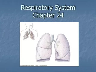
Chapter 14 - Respiratory System
260 likes | 300 Vues
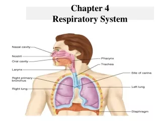
Chapter 14 - Respiratory System Ensures O2 gets to cells & CO2 removed from cells, helps maintain a constant environment in the body Organs - designed to perform 2 basic functions: Air Distributor - Gas Exchanger - Also -filters, warms, humidifies air

Chapter 14 - Respiratory System
E N D
Presentation Transcript
Chapter 14 - Respiratory System • Ensures O2 gets to cells & CO2 removed from cells, helps maintain a constant environment in the body • Organs - designed to perform 2 basic functions: • Air Distributor - Gas Exchanger - • Also -filters, warms, humidifies air • Sinuses - speech, sound, smell (olfaction)
Structural Plan - • Basic structure is like a many branched tube (respiratory tree) - nose, pharynx, larynx, trachea, bronchi, lungs • Alveoli - thin-walled sacs at end of tubes where gas is exchanged • Millions in each lung - > surface area • Each is covered with a network of capillaries (like a hairnet), also, wall is single layer thick • Gases (O2 & CO2) are exchanged by passive diffusion through the respiratory membrane (very thin - 1 micron thick)
Respiratory Tract - • Divided to assist in description of symptoms associated w/ problems • Upper - nose, pharynx, larynx (URI - head cold) • Located outside thoracic cavity • Lower - trachea, all parts of the bronchial tree, & lungs (LRI - chest cold) • Located inside thoracic cavity
Respiratory Mucosa - • Membranes that lines most of the distribution tubes • Air entering nose - contaminated w/ irritants (insects, dust, pollens, bacteria) • Mucous acts as most important air purification mechanism, traps almost everything < air to alveoli • 125ml mucous / day • Mucous blanket - continuous sheet • Moved upward to pharynx by cilia that cover epithelial cells (beat one way) [smoking-paralyze]
Nose - • External nares (nostrils) air enters & moves to R & L nasal cavities (lined w/ mucosa), parititioned by nasal septum • Surface - moist (mucous), warm (many blood vessels), olfactory receptors that are nerve endings (sense of smell) • Paranasal sinuses - continuous mucosa (4- frontal, maxillary, sphenoidal, ethmodial) all drain into the nasal cavities, hollow help lighten skull & serve as resonant chambers for production of sound
Lacrimal sacs - two ducts located inner aspect of eyes, drain tears into nasal cavity • Conchae (KONG-kee) - three shelf-like structures that protrude into the nasal cavity (both sides) • Mucosa-covered • > Surface area to warm and humidify • Supplemental O2 - bubbled thur water to humidify (if not dries & irritates resp. tract)
Pharynx - • Called the throat - about 5 inches long • 3 portions - • Nasopharynx - upper part, behind nose - contain auditory (eustachian) tubes which connect to middle ear (equalizes air pressure between middle & exterior ear), continuous mucosa (infections) • Oropharynx -behind mouth • Laryngopharynx - lowest portion • Serves as passage of air (shared w/ GI)
Tonsils - • Masses of lympathic tissue in pharynx • Pharyngeal tonsils - nasopharynx • Called adenoids if swollen, make breathing thru nose difficult • Palatine tonsils - oropharynx • Tonsillectomy - removal of tonsils • Tonsillitis - inflammation
Larynx - voice box (just below pharynx) • Made of cartilage (largest - thyroid cartilage - Adam’s apple) • Vocal cords - 2 fibrous bands, stretch across interior of larynx • Muscles cause them to tense (high pitched) & relax (low pitched) • Glottis - space between vocal cords • Epiglottis - cartilage, partially covers the opening of larynx (trapdoor), closed when swallowing
Trachea - • Windpipe - 4 1/2 inches long • From larynx to bronchi • Lined with mucous membrane • Vital function - furnishes open airway • Considerable force to squeeze closed • Almost Noncollapsible material - 15 to 20 C-shaped cartilage, stacked (soft tissue between) • Obstructed by: tumors, enlarged lymph nodes, aspirate food (choking-5th leading cause of accidental death in US)- Heimlich Maneuver - open windpipe that is suddenly obstructed
Bronchi, Bronchioles, & Alveoli - • Upside down tree (bronchial tree) • Larger tubes are ringed with cartilage • Primary bronchi -R & L >R & L lungs • Secondary bronchi - branches in each lung • Divide into smaller & smaller tubes - ultimately into tiny tubes made of smooth muscle - bronchioles > divide into microscopic tubes - alveolar ducts end in several alveolar sacs (cluster of grapes)
Alveolar sacs - (cluster of grapes) - each cluster made up of numerous alveoli (single grape) • Alveoli - very effective in gas exchange • Thin walled, each in contact w/ blood capillary • Surfactant - substance that covers the resp. membrane in the alveoli • Helps reduce surface tension > keeps alveoli from collapsing as air moves in & out
IRDS (Infant resp. distress syndrome) - • Premature infants (< 37 weeks gestation or wt. < 5 lbs. at birth), sacs collapse during expiration - inspiration requires more force to reinflate > labored breathing • Lungs & Pleura – • R - three lobes L - two lobes • Apex - upper end toward collarbone • Base - of lungs rest on diaphragm
Pleura - serous membrane linings, thin, moist, slippery • Parietal pleura - lines walls of thorax • Visceral pleura - covers the lungs • Intrapleural space - between , small amt. fluid • Pleurisy - inflammation of parietal pleural • Difficulty breathing, stabbing pain (rubs) • Caused by infection, tumors, etc. • Atelectasis - collapse of the lung > effective breathing due to < ventilation • Pneumothorax - Hemothorax
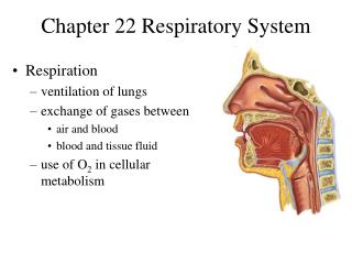
Respiration - • Exchange of gases between living organism & environment - Pair of lungs - place where air & circulating fluids (blood) can exchange gases • Pulmonary ventilation - (breathing) air in & out of lungs (external respiration- exchange gases) • Internal respiration - exchange gases between blood & cells • Cellular respiration - use of O2 by cells • Mechanisms of Breathing - Pulmonary Ventilation - 2 phases (inspiration - expiration) • Changes in pressure cause movement of air in-out
Lungs inside thoracic cavity - changes in shape & size of cavity (by muscles)- changes air pressure w/in cavity • Air moves from area of high pressure to area of low pressure • Inspiration - occurs when chest cavity enlarges > lungs expand > air rushes in & down to alveoli • Inspiratory muscles - Diaphragm & external intercostals • When these muscles contract > ^ volume in cavity > which < pressure - draws air in
Diaphragm - • Most important muscle of inspiration • Dome-shaped muscle • Flattens (contract) during inspiration > cavity elongate (top to bottom) • Phrenic nerve - stimulates to contract • External Intercostal - • Between the ribs • When contract > enlarge cavity (front to back) ( side to side) > < pressure (air rushes in)
Expiration - ordinarily passive process (quiet expiration) • Begins as inspiratory muscles relax • Cavity returns to smaller size • Lungs recoil - elastic nature < in size as air leaves • Expiratory muscles - used when speaking, sing, do heavy work • Need more forceful expiration to > depth & rate of ventilation • Internal intercostals & abdominal muscles • Cavity size < & pressure w/in > & air flows out
Internal Intercostals Muscles- • Depress the rib cage < cavity size front - back • Abdominal Muscles - • Contract & push abd. organs against underside of diaphragm(^ dome-shape) • Shortens top to bottom thoracic size • Exchange of Gases in Lungs - • Blood from R ventricle into pulmonary artery to lungs > tiny capillary beds close to alveoli • Diffusion occurs between tiny capillaries & alveoli (O2 & CO2)
Movement of substances from high concentration to area of low concentration • Oxyhemoglobin - O2 & hemoglobin combination in RBCs (to be carried to cells) • Most CO2 carried to lungs as bicarbonate ion (HCO3) , some carried in RBCs as carbaminohemoglobin • Exchange of Gases in Tissues - • Internal respiration - diffusion • Oxyhemoglobin breaks down > O2 into cells (used) - CO2 out of cells
Volumes of Air Exchanged in Pulmonary Ventilation - • Spirometer - device used to measure amt. air exchanged in breathing • Tidal Volume (TV) - (like ocean tides) amt. of air that comes & goes regularly • Normal Inspiration - 500 ml (one pint) • Normal Expiration - equal amt. • Vital Capacity (VC) - largest amt. air we can breathe out in one expiration • Normal young adult = 4800 ml
Expiratory Reserve Volume (ERV) - • Amount of air forcibly exhaled after tidal volume • Inspiratory Reserve Volume (IRV) - • Amount of air forcibly inspired over & above a normal resp. • As TV (normal breath) >, ERV & IRV < (reserve spaces) • VC = TV + IRV + ERV • Residual Volume (RV) - Air that remains in lungs after most forceful expiration
Regulation of Respiration - • Need for O2 > as activity > (make more waste products > removed) • Take more breaths (^ rate) & > tidal volume (depth) • Automatic adjustments - in resp. & circulation (heart pumps faster & harder) • Respiratory control centers - medulla & pons of brain - they stimulate resp. muscles • Receptors sense: Changes in O2 & CO2 levels in blood, Acid levels, Amt. stretch in lungs > change resp. rate & depth
Cerebral Cortex - • Modifying effect on inspiratory & expiratory centers of the medulla • Voluntarily change pattern of breathing • Hold breath (swimming, speaking, eating) • As limits - resume breathing when our bodies need O2 or has too much CO2
Receptors Influencing Respiration - • Chemoreceptors -in carotid & aortic bodies • Sense > CO2, < O2, acid levels in blood • Send nerve impulses to resp. regulation centers • Pulmonary Stretch Receptors - • Located in lungs - throughout airways & alveoli • Impulses influence normal breathing pattern to protect from excess stretching (overinhalation) • When TV reached - stimulus sent to inhibit more inspiration
Types of Breathing - • Eupnea - normal resp. rate, unaware of breathing • Hypoventilation - slow & shallow • Hyperventilation - rapid & deep • Dyspnea - labored or difficult • Orthopnea - upright position • Apnea - breathing stops • Cheyne-Stokes Resp. -apnea & hyperventilation • Respiratory Arrest - failure to resume breathing after period of apnea