Download
1 / 41
410 likes | 435 Vues
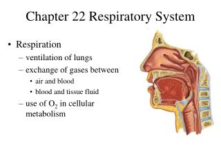
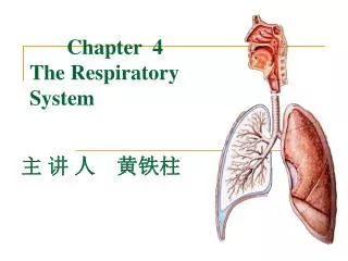
Chapter 4 Respiratory System. Respiratory System. Consists of the respiratory and conducting parts Respiratory part Site of gas exchange Consists of bronchioles, alveolar ducts, and alveoli Conducting part Provides rigid conduits for air to reach the sites of gas exchange

E N D
Respiratory System • Consists of the respiratory and conducting parts • Respiratory part • Site of gas exchange • Consists of bronchioles, alveolar ducts, and alveoli • Conductingpart • Provides rigid conduits for air to reach the sites of gas exchange • Includes all other respiratory structures (e.g., nose, nasal cavity, pharynx, trachea) • Respiratory muscles: diaphragm and other muscles that promote ventilation
Major Functions of the Respiratory System • To supply tissues with oxygen and dispose of carbon dioxide • Respiration: four distinct processes must happen • Pulmonary ventilation:moving air into and out of the lungs • External respiration: gas exchange between the lungs and the blood • Internal respiration: gas exchange between blood and tissues • Transport: transport of oxygen and carbon dioxide between the lungs and tissues
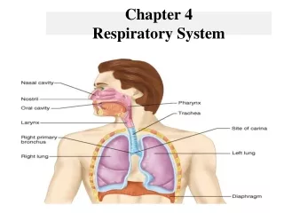
General Anatomy • Upper respiratory tract: • The nose, Mouth, pharynx, and Larynx. • Lower respiratory tract: • The trachea, Bronchi, Alveoli, and Lungs. • The thoracic cavity consists of: • Right and left pleural cavities (the parietal pleura lines the thoracic cavity, and the visceral pleura adheres directly to the lung tissue. • Mediastinum.
Respiratory Zone • Begins as terminal bronchioles, feed into respiratory bronchioles. • Respiratory bronchioles lead to alveolar ducts, then to terminal clusters of alveolar sacs composed of alveoli • Approximately 300 million alveoli: • Account for most of the lungs’ volume • Provide great surface area for gas exchange
Gross Anatomy of the Lungs • Lungs occupy all of the thoracic cavity except the mediastinum • Root or Hilum: site of vascular and bronchial attachments • Costal surface: anterior, lateral, and posterior surfaces in contact with the ribs • Apex: narrow superior tip • Base: inferior surface that rests on the diaphragm
Blood Supply to Lungs • Lungs are perfused by pulmonary and bronchial arteries. 1- Pulmonary arteries: • Supply systemic venous blood to be oxygenated • Ultimately feed into the pulmonary capillary network surrounding the alveoli • Pulmonary veins: carry oxygenated blood back to the heart 2- Bronchial arteries: • Provide systemic blood to the lung tissue • Arise from aorta and enter the lungs at the hilum. • Supply all lung tissue except the alveoli
Pleurae • Thin, double-layered serosa • Parietal pleura • Covers the thoracic wall and superior face of the diaphragm • Continues around heart and between lungs • Visceral pleura • Covers the external lung surface • Divides the thoracic cavity into three chambers • The central mediastinum • Two lateral compartments, each containing a lung
Mediastinum • Divided into: anterior, middle, and posterior portions. • Anterior mediastinum contains: the thyroid and thymus glands. • Middle mediastinum contains: heart and great vessels, esophagus, and trachea. • Posterior mediastinum contains: descending aorta and spine.
Imaging techniques used to investigate chest pathology include: • Plain X-Ray film • Computed Tomography (CT Scan) • Magnetic Resonance Imaging (MRI) • Ultrasound (US) • Angiography Chest X-Ray Review
Radiographic signs • Silhouette sign: opacity near the heart so you can not detect the heart boarder easily. • Notes from the radiographs: opacity frontal and heart frontal so lesion is frontal.
Negative Silhouette • Lesion Posterior.
Air Bronchogram • Opacity in the lung contains a tree filled with air (bronchi contains air but alveoli opaque). • Lesion arises from the lung not chest wall or pleura or mediastinum. • Air bronchogram=alveolar pathology=consolidation (lung filled with liquid).
Pulmonary nodule: well defined lesion less than 3cm. • Mass: well defined lesion more than 3cm.
To interpret chest x-raydifferentiate between • Focal lesion • Diffuse lesion
Focal lesions • Nodules • Masses • Cavities: well defined lesion (sphere-shape) filled totally with air or partially (air+liquid). • Patches: ill defined lesion (opacity) contains air bronchogram.
Nodules maybe due tomost common • Tuberculoma • Hamartoma • Bronchogenic carcinoma • Metastases (multiple nodules or solitary) • AVM (Arterio-Venus Malformation). • Hydatid cyst
Benign nodule (Hamartoma and Tuberculoma): If we have pulmonary nodule and the edge is smooth and contains calcification. A.V.M. bilateral
Lung Tubercloma: The term tuberculoma of the lung is used here to describe a rounded, homogeneous radiographic opacity, with well-defined borders, one centimetre or morein diameter.
How we can differentiate? • Tubercluma: usually single, smooth edge, calcified, less than 3 cm. • Hamartoma: usually single, smooth edge, popcorn calcification, less than 3 cm. • AVM: nodule connected to the hilum by feeding artery and draining vein.
Hamartoma: is a benign, focal malformation that resembles a neoplasm in the tissue of its origin. This is not a malignanttumor, and it grows at the same rate as the surrounding tissues. It is composed of tissue elements normally found at that site, but which are growing in a disorganized mass. They occur in many different parts of the body and are most often asymptomatic and undetected unless seen on an image taken for another reason • Lung Hamartoma: The most common hamartomas occur in the lungs. About 5–8% of all solitary lung nodules, about 75% of all benign lung tumors, are hamartomas.
Lung HamartomaBenign nodule is characterized by the presence of calcium but the presence of calcium in the mass can not be considered as a sign of benign lesion.
Bronchogenic carcinoma • Speculated margin nodule= malignancy. • Upper lobe distribution 70%.
Bronchogenic carcinoma • Lateral view showing the speculated margin.
Hydatid cyst • Single or multiple lesions filled with water. • Thin non enhancing margin. • Abscess: thick enhancing margin, usually contains air.
Patch • An ill defined lesion with air bronchogram. • Air bronchogram: air filled bronchi passing through opaque lung. • Pulmonary lesion. • Alveolar pathology. • Consolodation. • Patch may be due to: • Pneumonia or infarction.
Pneumonia • is an inflammatory condition of the lung affecting primarily alveoli. • It is usually caused by infection with viruses or bacteria. • Typical symptoms include a cough, chest pain, fever, and difficulty breathing
Diffuse lung disease • We have 4 radiographic pattern: • Reticular pattern e.g. Pneumonia, Bronchitis: Confluent small ill-defined densities produce a reticular pattern.
2. Ground glass pattern: increase in density in areas of ground glass and air trapping in lower lobes in patients with hypersensitivity pneumonitis (inflammation of lung tissue). Pneumonia is pneumonitis combined with consolidation (is a region of (normally compressible) lung tissue that has filled with liquid) due to infection.