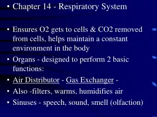
Respiratory System Chapter 24
Respiratory System Chapter 24. The main function of the respiratory system is to supply oxygen to, & eliminate carbon dioxide from the body In order to accomplish this task, the respiratory system must work in conjunction with the cardiovascular system.
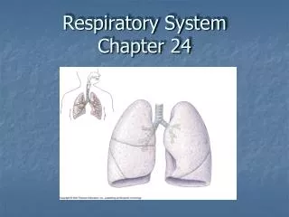

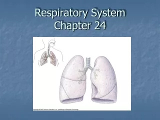
Respiratory System Chapter 24
E N D
Presentation Transcript
The main function of the respiratory system is to supply oxygen to, & eliminate carbon dioxide from the body In order to accomplish this task, the respiratory system must work in conjunction with the cardiovascular system
“Respiration” refers to the overall exchange of gases between the atmosphere, blood & cells • Respiration involves 3 processes • Pulmonary ventilation • External respiration • Internal respiration
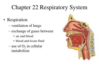
Anatomy Overview The respiratory tract includes: Nose (nasal cavity) Pharynx (nasopharynx, oropharynx, laryngopharynx) Larynx Trachea Bronchi (primary, secondary (lobar), tertiary (segmental) Bronchioles Terminal bronchioles Respiratory bronchioles Alveolar ducts Alveoli Nasal cavity Pharynx Larynx Trachea Bronchi Bronchioles Respiratory bronchioles Left Lung Right Lung Alveolar duct Alveoli
Histology Respiratory Epithelium = Pseudostratified Ciliated Columnar (PSCC)
Nose (nasal cavity) Air normally enters through external nares through nasal vestibule into nasal cavity. Nasal cavity divided by nasal septum and has a respiratory area with 3 nasal conchae (superior, middle & inferior) projecting into the midline from each lateral wall creating nasal meatuses between; and an olfactory area Nasal cavity communicates with the paranasal sinuses in frontal, maxillary, ethmoid & sphenoid bones Functions of nasal cavity include: warming, moistening & filtering air; olfaction
Pharynx Air passes from nasal cavity, across internal nares into nasopharynx, past oropharynx & through laryngopharynx to larynx Nasopharynx lined with PSCC epithelium, but oro & laryngopharynx lined with stratified squamous epithelium because they are also part of digestive system
Eustachian (auditory) tube – connects nasopharynx & middle ear cavity Pharyngeal tonsil – lymphatic tissue embedded in wall of nasopharynx Palatine tonsils - lymphatic tissue embedded in wall of oropharynx Uvula – posterior tissue from soft palate; protects nasopharynx when swallowing
Larynx Air passageway made of 9 pieces of cartilage – (1) Thyroid cartilage, (1) Epiglottis, (1) Cricoid cartilage, (2) Arytenoid, (2) Corniculate, (2) Cuneiform A.K.A your “voicebox” because it contains the vocal cords
Larynx • Thyroid cartilage – protects anterior & lateral walls of airway • Epiglottis – leaf-shaped cartilage that protects opening (“glottis”) of airway when swallowing • Cricoid cartilage – complete ring of cartilage; protects posterior wall of airway; attaches to trachea
Larynx • Arytenoid, corniculate & cuneiform cartilages – attach to upper (vestibular) vocal folds & lower (true) vocal cords
Trachea • Tough but flexible “windpipe”, anterior to esophagus • attached to cricoid cartilage (at about C6 vertebral level) & ends within mediastinum by branching into left & right primary bronchi (at T5 vertebral level) • End of trachea known as Carina Carina
Trachea • Lined with respiratory epithelium • “C”-shaped pieces of hyaline cartilage protecting airway while allowing for swallowing • Trachealis muscle (smooth muscle) runs across posterior wall of trachea connecting ends of tracheal cartilage
Trachea Low power High power Medium power
Bronchi • Trachea splits into a left & right primary bronchus which enters into the hilus of each lung • Within the lung, the primary bronchi branch into secondary (lobar) bronchi (3 in right lung/2 in left lung) • Secondary bronchi then branch into 10 tertiary (segmental) bronchi • Tertiary bronchi then continue to branch into smaller & smaller bronchi & then into very narrow bronchioles Carina This branching patterns creates the “bronchial tree”
Changes In Airway • As you go further down into the bronchial tree of each lung, changes in the airway occur: • increased number of airways (1 primary; 2 or 3 secondary; 10 tertiary bronchi; 6000 terminal bronchioles; millions of alveolar ducts) • decreased diameter of each airway • decreased amount of cartilage in the airways (no cartilage at all by terminal bronchioles) • increased amount of smooth muscle (relative to diameter) • lining epithelium changes from PSCC simple squamous epithelium (in alveoli)
Lungs Located within the thoracic cavity, surrounded by the double-layered pleural membrane – parietal pleura – lines cavity wall visceral pleura – covers the lungs
Apex – extends 1” above clavicle Hilum – at medial surface; where primary bronchus, pulmonary artery & veins enter/exit lung Superior lobe Superior lobe Horizontal fissure Oblique fissure Middle lobe Oblique fissure Cardiac notch Inferior lobe Inferior lobe Base – rests on diaphragm Lungs- Anatomical Features Right lung Left lung
Lung – medial surface Groove for aorta Hilum Cardiac notch of Lt. lung
Airways within Lungs • Each lung has a primarybronchus entering at the hilus • Each lobe of a lung has a secondary (a.k.a. lobar) bronchus • Lobes are functionally divided into bronchopulmonary segments & each segment has a tertiary (segmental) bronchus • Segments are functionally divided into many lobules & each lobule receives a terminal bronchiole
Relationship of Airways & Pulmonary Vessels • As airways branch within lungs, they are accompanied by branches of the pulmonary artery (carrying de-oxygenated blood into the lungs), & branches of the pulmonary veins (carrying oxygenated blood out of the lungs) • As the alveolar ducts expand to form alveoli, pulmonary arterioles will branch to form a network of pulmonary capillaries, surrounding the alveoli
Alveoli • Alveoli are expanded chambers of epithelial tissue that are the exchange surfaces of the lungs • There are about 150 million alveoli in each lung • Multiple alveoli usually share a common alveolar duct, creating “alveolar sacs”
There are three types of cells found within alveoli: • Alveolar Squamous epithelial (aka “type I”) cells – primary cells making up the wall of the alveoli • Septal (aka “type II”) cells – sectrete “surfactant” to reduce surface tension which prevents alveoli from sticking together & allows for easier gas exchange • Alveolar macrophages (aka “dust cells”) – phagocytic cells that remove dust, debris & pathogens Alveoli
Gas “exchange” occurs across the Respiratory membrane – the fused membranes of the alveolar epithelium & the pulmonary capillary endothelium