
Uploaded by
nadine
0 SLIDES
187 VUES
0LIKES
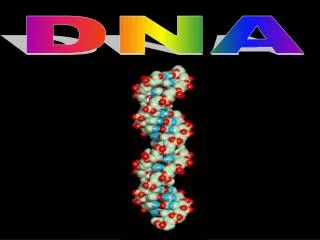
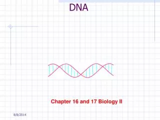
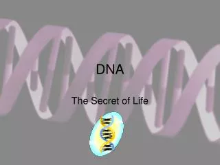
DNA
DESCRIPTION
DNA. EUKARYOTIC CELLS. SUPER COILS. CHROMATIN. DOUBLE HELIX COILS AROUND PROTEINS (HISTONES) HISTONES COIL MOLECULE INTO SUPER COILS SUPER COILS WIND UP TO FORM CHROMOSOMES CHROMOSOMES FORM CHROMATIN IN NUCLEUS. PRIMARY FUNCTION.
Download

1 / 0
Télécharger la présentation 
DNA
An Image/Link below is provided (as is) to download presentation
Download Policy: Content on the Website is provided to you AS IS for your information and personal use and may not be sold / licensed / shared on other websites without getting consent from its author.
Content is provided to you AS IS for your information and personal use only.
Download presentation by click this link.
While downloading, if for some reason you are not able to download a presentation, the publisher may have deleted the file from their server.
During download, if you can't get a presentation, the file might be deleted by the publisher.
E N D
Presentation Transcript
-
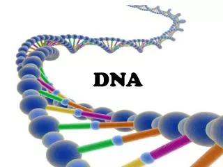
DNA
- EUKARYOTIC CELLS SUPER COILS CHROMATIN DOUBLE HELIX COILS AROUND PROTEINS (HISTONES) HISTONES COIL MOLECULE INTO SUPER COILS SUPER COILS WIND UP TO FORM CHROMOSOMES CHROMOSOMES FORM CHROMATIN IN NUCLEUS
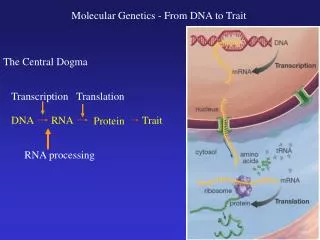
- PRIMARY FUNCTION STORES AND TRANSMITS GENETIC INFORMATION BASED UPON CODE SEQUENCE CHEMICALLY CONTROLS THE SYNTHESIS OF ALL PROTEINS ENZYMES THAT CONTROL CHEMICAL PROCESSES DETERMINE PHYSICAL STRUCTURES OF ORGANISMS
- DeoxyriboNucleicAcid Made of repeating monomers: NUCLEOTIDES 5-carbon sugar Phosphate functional group 1 of 4 Nitrogen containing bases Adenine Cytosine Guanine Thymine
- NITROGEN BASES PURINES PYRIMIDINES BASES WITH ONE CARBON RING Uracil only found in RNA BASES WITH TWO CARBON RINGS
- BASE PAIR BONDING ONE PURINE BONDS WITH ONE PYRIMIDINE FORMING A COMPLIMENTARY BASE PAIR ADENINE ALWAYS BONDS WITH THYMINE FORMING A-T OR T-A GUANINE ALWAYS BONDS WITH CYTOSINE FORMING G-CORC-G
- THAT’S INTERESTING! DNA LOOKS LIKE A LADDER SUGAR AND PHOSPHATE MAKES THE “FRAME” BASES FORM THE “RUNGS” ONE SIDE OF MOLECULE MUST “FLIP” IN ORDER FOR HYDROGEN BONDS TO FORM BETWEEN BASES, IT IS ANTIPARALlEL ALL BASE PAIRS, NO MATTER THE ORDER, HAVE THE SAME STRUCTURAL SHAPE SO THEY STACK ON TOP OF EACH OTHER THEN TWIST FORMING THE CHARACTERISTIC DOUBLE HELIX SINCE A ALWAYS BONDS WITH T, AND C WITH G, THEIR AMOUNTS MUST BE EQUAL TO EACH OTHER(Chargaff Principle) NATURE LOVES EFFICIENCY! DNA’S STRUCTURE ALLOWS THE MOLECULE TO REPLICATE ITSELF QUICKLY, AS WELL AS, CODE FOR THE SYNTHESIS OF PROTEINS WITHIN THE ORGANISM(ALL ORGANISMS), AND PASS THE GENETIC CODE TO FUTURE GENERATIONS; THE SAME WAY EVERY SINGLE TIME.
- Who figured all this out? Rosalind Franklin and Maurice Wilkins Maurice Wilkins and Rosalind Franklin were the first to obtain very good x-ray diffraction images of the DNA fibers. At that time, little was known about the structure of DNA; though these photos didn't show the structure of the DNA, there were patterns on those images that could be used to determine the position of the DNA molecule's atoms. From these photos, Franklin determined that the DNA molecule must be long and thinand possibly a helix
- James Watson and Francis Crick In 1951 James Watson and Francis Crick began to examine the DNA’s structure. Using previous X-ray diffraction photos of DNA fibers taken by Maurice Wilkins and Rosalind Franklin, they discovered that it showed an X shape... which is also the characteristic of a helix. In April of 1953, using this information, together with the base pair’s isometric structure, they identified DNA’s double helixconfiguration.
- DNA Replication - Occurs during S phase of interphase One strand of DNA has all of the information needed to reconstruct the other half through the mechanism of base pairing. During DNA replication, the DNA molecule is used to produce two new complementary strands. Each strand of the double helix serves as a template for the new strand.
- Old Model of DNA Replication Enzyme unzipped molecule down entire length Enzymes attach to each side Enzyme brings in complimentary bases New strands build simultaneously in same direction until duplicate strands are formed
- DNA Replication Step 1: A portion of the parental DNA double helix is unwound and separated by the enzyme helicase. Step 2: Proteins attach to outside of replication fork to stabilize the molecule
- DNA Replication Step 3: The enzyme DNA polymerase attaches to one strand of the DNA and begins synthesis of the leading strand. Because sugars are right side up…this strand is replicated continuously
- DNA Replication Step 4: Because the other side of the molecule is upside down, and the enzymes only work in one direction, this side has to be replicated “discontinuously” in sections. BANANA
- DNA Replication DNA replication is “semiconservative”. This means that one-half of each new molecule of DNA is old; one-half new.


More Related