Download
1 / 77
770 likes | 1.1k Vues
Biological effects of ionizing radiations. DNA structure and function Molecular organization of life DNA structure and function Diversity and function of biological macromolecules. Dosimetry : Absorbed dose Equivalent and effective dose The W R and W T coefficients.

E N D
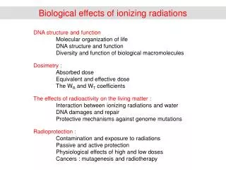
Biological effects of ionizing radiations DNA structure and function Molecular organization of life DNA structure and function Diversity and function of biological macromolecules Dosimetry : Absorbed dose Equivalent and effective dose The WR and WT coefficients The effects of radioactivity on the living matter : Interaction between ionizing radiations and water DNA damages and repair Protective mechanisms against genome mutations Radioprotection : Contamination and exposure to radiations Passive and active protection Physiological effects of high and low doses Cancers : mutagenesis and radiotherapy
Organization levels in biology Small molecules Biological macromolecules Macromolecules nm Molecular assemblies Energy flow Genome Intracellular compartments mm Cells Plasma membrane Organisms mm Population
Information flow in living cells promoter DNA (desoxyribonucleic acid) mRNA (ribonucleic acid) proteins enzymes transcription factors plasma membrane receptors small molecules
DNA structure 3 ’ 5 ’ base 1 base 3 3,4 Å 34 Å base 2 base 2 20 Å sequence base 3 base 1 5 ’ 3 ’ Direct strand Complementary strand
Interaction between DNA strands Adenine thymine pair Complementary strands are opposed Guanine-cytosine pair
DNA is organized in chromosomes procaryotes : a single circular chromosome typically 5.106 base pairs eucaryotes : several linear chromosomes typically 3.109 base pairs telomer centromer 22 autosomal chromosome pairs Example : human genome + about hundred circular mitochondrial DNA molecules 2 sexual chromosomes
DNA, RNA and proteins DNA-dependant RNA polymerase nucleotides TRANSCRIPTION Transfert RNA (tRNA) aminoacyl tRNA transferase ribosomes TRANSLATION -COO- NH3+- -COO- NH3+-
A G U/T C The genetic code Three consecutive nucleotides form a codon that encodes an amino acid U/T C A G U/T U/T C A G C Genetic engineering : production of new proteins, using modified DNA U/T C A G A START U/T C A G G
The various sets of biological molecules DNA replication and repair genome DNA noncoding RNA and mRNA synthesis mRNA editing and degradation transcriptome (m)RNA protein synthesis Protein post-translational modifications proteome proteins protein activity Protein degradation small molecules metabolome
Molecular diversity in different model organisms Saccharomyces cerevisiae Escherichia coli Homo sapiens 12 Genome size 5.106 3.109 Number of genes 4639 6607 ≈ 25000 Number of proteins 4289 6300 ≈106 Number of cell types 2 2 ≈ 250 Number of cells 1-109 1-108 1014 Cell size 1-2 mm 5 mm 10-50 mm
ENCODE project (summer 2012) : a function can be attributed to 75% of the human genome
Biological effects of ionizing radiations DNA structure and function Molecular organization of life DNA structure and function Diversity and function of biological macromolecules Dosimetry : Absorbed dose Equivalent and effective dose The WR and WT coefficients The effects of radioactivity on the living matter : Interaction between ionizing radiations and water DNA damages and repair Protective mechanisms against genome mutations Radioprotection : Contamination and exposure to radiations Passive and active protection Physiological effects of high and low doses Cancers : mutagenesis and radiotherapy
From the absorbed radiation dose to the equivalent and the effective doses physico-chemical effects biological effects ionizing radiation exposure absorbed radiation dose equivalent dose effective dose absorbance + diffusion ionization cell death cancer risk C/kg J/kg = gray sievert sievert (röngten) (rad) (rem) (rem) Real-time measurements possible Delayed and multiple effects http://www.euronuclear.org/info/
Measuring the exposure to ionizing radiations Unit : C/kg 1 C/kg is the amount of radiation required to create 1 C of positive and negative electrostatic charges in 1 kg of matter. This corresponds to the generation of 6.1018 ion pairs. Old unit : röentgen 1 R is the amount of radiation required to create 1 C of positive and negative electrostatic charges in 1 cm3 of dry air at 20°C and 1 atm. 1 R = = 2.58×10⁻⁴ C/kg Measurement : Geiger and scintillation counters
Measuring the absorbed radiation dose Unit : Gray (J/kg) The amount of radiation energy adsorbed per mass unit 1 Gy = 1J/kg = 1 N.m/(N/(m.s-2)) = 1 m2.s-2 Old unit : rad = roentgen absorbed dose 1 rd = 10-2 Gy ( = 100 erg/g) Measurement : passive and active dosimeters Note : doses can also be calculated from the geometry of the beams, the radioactive decay rate (radionucleid activity) and the composition and shape of the irradiated material
Passive dosimeter (dose integrated over time) thermoluminescent dosimeter film dosimeters Differential sensitivity to radiations : open window : b particles plastic : a particles copper filter : g photons aluminium (cadmium) filter : g photons > 150 keV, a particles > 2 MeV
Active dosimeter (dose rate) electronic dosimeters Principle Scintillation crystals and solid state detector Specifications Sensitive to X and μ radiation, ß particles Neutron response <2% Dose display and storage 0 μSv to >16 Sv Dose rate display 0 μSv/h to >4 Sv/h
Calculating the equivalent and the effective radiation doses The biological effect of radiations depends on : The type of the radiation. It is greater for radiations that have a high ionization density along the track of particles (linear energy transfert LET). The cell type. It is greater for cells that divide rapidly, cells that undergo DNA recombination and stem cells. Equivalent radiation dose = absorbed radiation dose x radiation weighting factor (Sv) wR (quality factor, no unit) Effective radiation dose = equivalent radiation dose x biological weighting factor (Sv) wT (weighting factor, no unit) For several radiation types and for several cell types, contributions are linearly summed up :
wR factors particles/radiations energy wR photons g all 1 a particles all 20 b particles all 1 protons < 10 keV 5 10 -100 keV 10 > 20 MeV 5 neutrons 100 keV - 2 MeV 20 2 - 20 MeV 10 > 20 MeV 5 Photons, a and b particles : ICRP 60 (1990) Protons and neutrons : ICRP 92 (2003) ICRP : international commission on radiological protection CIPR : commission internationale de protection radiologique These values are based on cell death measurements
wT factors Cell type wT(1) wT(2) gonads (gamet organs) 0.20 0.08 bone marrow 0.12 0.12 colon 0.12 0.12 lung 0.12 0.12 stomach 0.12 0.12 bladder 0.05 0.04 breast 0.05 0.12 liver 0.05 0.04 thyroid gland 0.05 0.04 skin 0.01 0.01 bone surface 0.01 0.01 brain 0.01 S wT = 1 body (1) ICRP 60 (1990) (2) ICRP 92 (2003) ICRP : international commission on radiological protection CIPR : commission internationale de protection radiologique These values are based on fatal cancer occurrence Robert N. Cherry ENCYCLOPÉDIE DE SÉCURITÉ ET DE SANTÉ AU TRAVAIL, les rayonnements ionisants
Biological effects of ionizing radiations DNA structure and function Molecular organization of life DNA structure and function Diversity and function of biological macromolecules Dosimetry : Absorbed dose Equivalent and effective dose The WR and WT coefficients The effects of radioactivity on the living matter : Interaction between ionizing radiations and water DNA damages and repair Protective mechanisms against genome mutations Radioprotection : Contamination and exposure to radiations Passive and active protection Physiological effects of high and low doses Cancers : mutagenesis and radiotherapy
Explaining WR, the dependence of the biological effects on the radiation type Most damages are due to water radiolysis, that generates reactive oxygen species The reactive oxygen species diffuse about 5 µm away from the ionizing particle/radiation The linear energy transfer strongly depends on radiation type
Most DNA damages are indirect Macromolecule damages induced by ionizing radiations cell fraction (w/w) molecule damage induced by radiation 70% H2O reactive ion production adducts, breaks → mutagenesis 1% DNA 5% lipids cell lysis 20% proteins inactivation or spontaneous activation of metabolic or signaling pathways <1% mRNA
Water radiolysis : end products H2O* H2O e-aq + H2O+ OH- hydroxide ion detoxification enzymes H2O2 hydrogen peroxide peroxidase O2- superoxide ion superoxide dismutase (SOD) Reactive Oxygen Species (ROS) Dissolved oxygen promotes the formation of reactive oxygen species
Water radiolysis : detailed mechanisms Diffusion typically 5 µm strong reducing agents strong oxidizing agents strong oxidizing agents Sophie Le Caër (2011) Water Radiolysis: Influence of Oxide Surfaces on H2 Production under Ionizing Radiation. Water 3, 235-253
O H O H O O O O H O O Water radiolysis products A radical is a chemical (atom, ion, molecule) with unpaired electrons on the highest energy orbital. Radicals are often very reactive. OH- : hydroxyl ion OH : hydroxyl radical O2- : superoxide ion HO2- : hydroperoxyl radical 02 : dioxygen (a stable radical)
Linear Energy Transfert (LET) L (J.m-1): linear energy transfert Energy decrease per unit length
Linear Energy Transfert (LET) and WR coefficients Unlimited linear energy transfer L in water (keV.µm-1) Q(L) < 10 1 10 – 100 0.32 L – 2.2 > 100 300.(L)-0.5 http://www.euronuclear.org/info/encyclopedia/q/quality-factor.htm L = 5300 keV/37 µm = 143 WR = 25
The Bragg peak For fast moving particles and ions, most of the dose is deposited at the end of the particle track
Application to radiotherapy g photons all organs a, b particles internal exposure, skin Protons, neutrons, carbon ions variable deepness (Bragg peak)
Explaining WT, the dependence of the biological effect on the type of tissue/organ • There are three levels of cell protection against DNA damages • Scavenging DNA damaging molecules, especially reactive oxygen species • DNA repair mechanisms • Cell cycle checkpoints and apoptosis. Too many DNA damage leads to programmed cell death • Cell death induced by Natural Killer Cells and cytotoxic T cells from the immune system (detect changes in protein expression)
1. Superoxide dismutase and hydrogen peroxidase 2 O2− + 2 H2O O2 + H2O2 + 2 OH− HOOH + electron donor (2 e-) + 2H+ 2H2O Human mitochondrial SOD Mn cofactor PDB 1VAR 1 nm Bovine gluthatione peroxidase PDB 1GP1 8 genes in humans expressed in different tissues + 6 peroxiredoxins + catalase 3 genes in humans coding for extracellular, cytoplasmic, and mitochondrial isoforms
2. Sources of DNA damage Replication errors: DNA polymerase frequency 1/107 Molecular damages to DNA: Origin DNA damage number/cell.day Possible repair Exogenous sun (1h/day) T-T dimers 6-8.104 Y chemical adducts 102-105N (base modification) radioactivity single strand breaks 2-4.104 Y (natural double strand breaks ? ± background) Endogenous temperature single strand breaks 2-4.104 Y free radicals adducts/breaks 104 Y metabolites adducts 102 Y viruses genome integration ? N transposons ? ?
DNA repair mechanisms Damage type Repair • Recognition T-T dimers Adducts Single strand breaks Double strand breaks • Restriction • Excision • Synthesis • Ligation • Excision • or direct ligation • Recombination • Ligation
The COMET assay to measure DNA damages also called single cell gel electrophoresis (SCGE)
Exemple of repair : thymine dimers (induced by UV light) Tymine dimer repair enzyme : specific DNA endonuclease
translocation to the nucleus aromatic molecule (L) Aryl hydrocarbon Receptor AhR AhR-L AhR-L AhRE AhRE induction of specific mRNA (AhRE) P450 cytochromes (phase I) : CYP1A1, CYP1A2, CYP1B1, CYP2S1 Phase II enzymes : GST, UGT (detoxification mechanism) Growth Differentiation Metabolism (toxicity) CYP1A1, CYP1A2 epoxide hydrolase the diol epoxide covalently binds to DNA (adduct) Increased DNA mutations & cancer benzo[a]pyrene-7,8-dihydrodiol -9,10-epoxide Metabolism et carcinogenicity of Benzo[a]Pyrene Benzo[a]pyrene is a product of incomplete combustion at temperatures between 300 and 600 °C. benzo[a]pyrene (BP)
Shimizu et al. (2000) PNAS 97 : 779-782 Benzo[a]pyrene carcinogenicity is lost in mice lacking the aryl hydrocarbon receptor Individual susceptibility to xenobiotics. Exemple of CYP genes Dossier INSERM Dioxines dans l’environnement. Quels risques pour la santé? http://ist.inserm.fr/basisrapports/rapport.html
Indirect carcenogenicity of dioxin 2,3,7,8-tetrachlorodibenzo-p-dioxin (TCDD) Travailleurs exposés aux phénoxy-herbicides et aux chlorophénols. Exposition : 3 à 389 pg/g de matières grasses Teneur du lait maternel en France : 16,5 ± 5 pg/g de matières grasses Dossier INSERM Dioxines dans l’environnement. Dioxins occur as by-products in the manufacture of organochlorides, in the incineration of chlorine-containing substances such as PVC, in the bleaching of paper, and from natural sources such as volcanoes and forest fires. Dioxins build up primarily in fatty tissues over time. The major source of dioxins is food, especially from animals. TCDD has a half-life of approximately 8 years in humans. TCDD activates the AhR and thus induces CYP expression. This either increases or reduces carcinogenicity of other aromatic molecules such as Benzo[a]Pyrene and 7,12-dimethylbenz[a]anthracene, respectively.
The rad genes in yeast • A systematic study was conducted in yeast to identify genes responsible for the cell sensitivity to radiation 55 “rad” genes were found. • Most of these genes have counterparts in the human genome. • From current estimates, 240 genes are involved in DNA repair in humans
P Perego (2000) Yeast Mutants As a Model System for Identification of Determinants of Chemosensitivity. Pharmacol Rev52: 477–491
3. The cell cycle G0 Mitosis Gap 2 In the resting state (G0), cells don’t divide Gap 1 DNA Synthesis In tissues, most cells are in the resting state. Division occurs to self repair the tissue See ‘wound repair’ and ‘breast cancer cells’ movies
p53 Retinoblastoma protein (Rb) Anaphase Promoting Complex (APC) APOPTOSIS APOPTOSIS APOPTOSIS Cell cycle checkpoints Apoptosis is an organized (programmed) cell death mechanism
Apoptosis • Apoptosis is one form of programmed cell death, often observed in higher eukaryotes during development, selection of immune system cells, and cancer prevention by NKC • Apoptosis can be triggered by intracellular processes, such as DNA damages, or by extracellular molecules, for instance activation of the Fas receptor by the Fas ligand, or the secretion of permeabilizing molecules by NKC. • Apoptosis involves mitochondrial inactivation and the release of cytochrome c in the cytosol. • The lack of ATP induces phosphatidyl serine exposure to the plasma membrane (the “eat-me” signal) and cell blebbing. • Cell fragments are internalized by macrophages and digested. No inflammation (activation of the innate immune system) occurs . See movie 18.1 apoptosis
The p53 protein holds the cell cycle at the G1/S checkpoint in the presence of DNA damage p53 is a tetrameric 393 aa protein p53 consists of 3 domains : 1 100 200 300 393 phosphorylations NLS acetylations transcription activation domain DNA binding domain regulatory domain The transcription activation domain interacts with the Mdm2 protein that triggers p53 degradation. The DNA binding domain interacts with a specific DNA sequence that controls p21CIP expression The conformation and the localization of p53 is controlled by phosphorylation and acetylation p53 DNA binding domains in complex with DNA
ADN intact damaged Chk inactive active p53 absent bound to DNA (mdm2) (phosphorylated) p21 repressed expressed CDK active inactive Cycle G1S G1 stop p21 TRANSCRIPTION of p21 inhibitor Mdm2 p53 p53-Ub synthesis degradation + Chk1/2 + DNA polymerase p53-P Double strand break Single strand break (30 to 40 bases lacking) cycline E Base mispairing P cdk2 Mdm2 = murine double minute oncogene Chk = checkpoint kinase p21= CIP (cdk2 inhibiting protein) = WAF1 (Wild Type p53-activated fragment)
p53 mutations are found in 50% of human cancers Arg175, 249, 273, 282, Gly245 Arg248 Mutation frequency Séquence primaire de p53 • These mutations decrease p53 interaction with DNA, which eliminates the G1/S restriction point controlled by the Cdk2-cyclinE complex
4. Natural Killer Cells and cancer prevention • Natural Killer Cells (NKC) are components of the (innate) immune systems. They are cytotoxic against tumor cells and cells infected by viruses. They also play an important role in graft rejection. • NKC are sensitive to the molecules present at the surface of the cells. All cells in the body express MHC-I complexes that present fragments of endogenous proteins synthesized in the cell. Any change in the nature of MHC-I or in the surface concentration of MHC-I leads to NKC activation • Upon activation, NKC bind to the target cell and locally release perforin and granzyme molecules at the plasma membrane of the target cell, which triggers apoptosis. • In addition, NKC are able to recognize and kill cells with antibodies bound at their surface (adaptative immune system). Antibodies directed against surface antigens are indeed often present in cancers. • Defects in NKC production severely increases the risk of cancer • Tumor cells develop inhibitors that prevent NKC activation See movie 24.4 killer T cells
DNA repair mechanisms and cell fate DNA (desoxyribonucleic acid) mRNA (ribonucleic acid) UNCONTROLLED CELL DIVISION = CANCER proteins Repair enzymes Transcription factors CELL DIVISION Replication factors DNA state sensors Apoptosis factors (mitochondrial inactivation, caspases) CELL APOPTOSIS = CELL DEATH p53 Radiations DNA damage apoptosis Rapidly dividing cells are more sensitive to radiations
Molecular origins of cancer and possible molecular therapies oxidative stress xenobiotics radiations viruses & transposons detoxification (AhR, CYP) tanning immune system DNA damage DNA integration DNA repair (Xp) cell apoptosis cell killing (NK cells, cytotoxic T cells) permanent mutations permanent insertions DNA damage monitoring (p53, Mdm2) Cell cycle checkpoints (Retinoblastoma) Response to growth factors (RTK, Ras, CTK, etc…) accelerated mutagenesis and clonal selection escaping the immune system proliferation metastasis & angiogenesis radiotherapy cell division inhibitors (taxol) growth inhibition (tamoxifen) angiogenesis inhibitors (angiostatin) immunotherapy apoptosis tumor dormancy cell killing