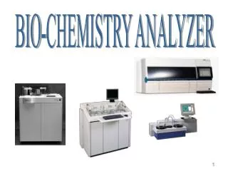
BIO-CHEMISTRY ANALYZER
BIO-CHEMISTRY ANALYZER. DIRECTORATE OF BIOMEDICAL ENGINEERING. PREPARED & PRESENTED BY. Eng . Khaled Masalha. Eng . Anton Khleif. Contents. Principals of Spectrum Beer’s Law Block diagram Sample reading Chemistry block diagram Measurement Principles of Chemistry analyzer

BIO-CHEMISTRY ANALYZER
E N D
Presentation Transcript
DIRECTORATE OF BIOMEDICAL ENGINEERING PREPARED & PRESENTED BY Eng . Khaled Masalha Eng . Anton Khleif
Contents Principals of Spectrum Beer’s Law Block diagram Sample reading Chemistry block diagram Measurement Principles of Chemistry analyzer Photometry Analyzer Photometric Measurements Kinetic or Enzymatic Measurements End Point Procedures Application Units BASIC ISE ISE Theory ANALYTICAL MEHOTDS SAFETY REGULATIONS Trouble shooting
Principals of Spectrum Many compounds absorb ultraviolet (UV) or visible (Vis.) light. The diagram below shows a beam of monochromatic radiation of radiant power P0, directed at a sample solution. Absorption takes place and the beam of radiation leaving the sample has radiant power P.
The amount of radiation absorbed may be measured in a number of ways: Transmittance, T = P / P0% Transmittance, %T = 100 TAbsorbance, A = log10P0/ PA = log10 1 / TA = log10 100 / %TA = 2 - log10 %T The last equationallows you to easily calculate absorbance from percentage transmittance data. Beer’s Law
The Beer-Lambert Law the equation representing the law is : A=ebc Where A is absorbance (no units, since A = log10P0/ P ) e is the molar absorbtivity with units of L mol-1 cm-1 b is the path length of the sample - that is, the path length of the cuvette in which the sample is contained. We will express this measurement in centimetres. c is the concentration of the compound in solution, expressed in mol L-1
The ABS Photometer is an opto-electromechanical module which performs light-absorbance measurements on samples. Light from its standard source passes through the cuvette. Certain wavelengths of the light are absorbed in varying degrees depending on the composition of the sample in the cuvette. These wavelength absorptions are measured to provide an analysis of the cuvette contents.
Absorbance Photometry is a measurement technique for determining concentrations of substances in fluid samples. Substances, or substances reacted with reagents, absorb specific wavelengths of light. The amount of absorbance is a measure of the concentration in solution. In the 8-12 wavelengths are available of which one or two are selected, the selection depending on the substance being measured. The 8-12 wavelengths are in the range from 340 to 660 nanometers (nm) as well as one (800nm) in the infrared range (visible light ranges from approximately 400 to 700nm). A software algorithm calculates the concentration of the substance in solution depending on the measured absorbance characteristics.
Sample reading Method for determination of glucose in whole blood and cuvette and photometer for carrying out said method A sample of whole blood is contacted with a reagent which by chemical reaction with glucose in the sample brings about a detectable dye concentration change the size of which is determined as a measure of the glucose content of the sample. The sample is initially introduced undiluted in a microcuvette having at least one cavity for receiving the sample. The cavity is internally pretreated with the reagent in dry form, and the chemical reaction takes place in the cavity. Active components of the reagent comprise at least a hemolyzing agent for exposing glucose contained in the blood cells of the sample for allowing total glucose determination, and agents taking part in the chemical reaction and ensuring that the dye concentration change takes place at least in a wavelength range outside the absorption range of the blood hemoglobin. An absorption measurement is performed in said wavelength range directly on the sample in the cuvette.
Measurement Principles of CHEMISTRY ANALYZER Modern medicine relies heavily upon the analysis of body substances to evaluate and confirm a diagnosis. These analyses allow physicians: - to prescribe a course of treatment for patients- to ascertain the stage of an illness and thus specify optimum treatments. Various body fluids may be analysed in different ways. Blood (both serum and plasma) and urine are the most commonly analysed, with blood being by far the more important of the two because of its various components.
Methods of analysis also differ widely, depending on purpose. The most useful and thus most commonly used areIonSelective Electrode (ISE)analysis andPhotometer analysis: these are used in the Instrument. There aretwo measurement areas in the Instrument: - ISE Module- Photometry Analyzer.
Photometry AnalyzerPhotometer ABS Absorption Analysis Photometry analysis methods are based upon measuring the amount of light absorbed by a substance. The measurements may be done directly on the substance or immediately after a chemical or enzymatic reaction. Chemical or enzymatic reaction methods require a reagent to be mixed with the sample before beginning the tests.
In certain situations, the concentration of a particular substance in a sample could be established by direct measurement of the amount of light absorbed. More often, however, the light absorption properties of a substance are inadequate for analysis purposes because interfering elements in the samples make them opaque. Thus samples normally are first mixed with a reagent (or sometimes two or three in sequence). The reagents are targeted to react with the substance to be measured only.
If the reagent is a chemical, the reaction converts the substance (or the reagent) into a specific quantity of dye product which can then be measured. This method may be used for defining most biologically significant metabolites in solution The concentration of a metabolite in solution may also be established using enzymatic techniques. In this method the enzyme converts a substance into a defined quantity of another product, which can be easily measured using photometric methods. This method is often the only one possible for complex biological mixtures. In a similar way enzymes in a sample can also be measured. Tracking enzyme levels in body fluids is an important diagnostic tool in modern medicine
Photometric Measurements The normal sequence for a photometric measurement procedure consists of: • Transfer (pipette) a known quantity of sample into a cuvette • Transfer appropriate reagent into the cuvette • Ensure that sample and reagent are thoroughly mixed • Add and thoroughly mix any additional (e.g., starting) reagents required. • Carry out photometric measurements • Calculate and evaluate results
Concentration levels are calculated using the formula: Concentration = Absorption x Factor The multiplying factor is dimensioned depending on: - Wither the absorption is at the absolute level of absorption- The change in absorption between the start and end of the test- The rate of change in absorption with respect to time.
Calculation of a factor may be based on information provided on the Reagent. Alternately, it may be determined by carrying out one or more calibration runs using a calibrator or a standard substance instead of a real sample. In such situations, the calibration run is repeated periodically (e.g. every five hours or at beginning of day [BOD]), or when a new Reagent is used. A calibrator is a serum-based substance that is used for calibrating several points for differing analysis tests.
Kinetic or Enzymatic Measurements Kinetic or enzymatic Measurement procedures involve repeated photometric measurements at defined time intervals. For these measurements (also called continuous measurements), measurement values are obtained from a series of known standards and plotted using an x-y coordinate system. Each value represents a specific point in time (x axis) versus its corresponding absorbance value (y axis). Connecting the points producesa curve.
Values obtained from patient samples are compared against the "standard" curve to determine the concentration of enzyme in the sample. This curve may not necessarily be linear. The results may also be distributed. In some cases the actual change in value of absorption is known (i.e. the operator enters the measurement range). It is then only necessary to measure the time for the absorption to change from one value to the other. This provides the rate of change associated with the particular sample. Alternatively, it may be necessary to evaluate the slope of the curve where it is most linear. Here a linear regression process is applied to all data points to establish the required value of change in absorption with respect to change in time.
End Point Procedures End point procedures look for a value at the end of a test. The rate of change in absorption with respect to time may be variable from test to test and therefore not suitable for measurement purposes. In this case it may be possible to establish the level of concentration of a substance by measuring the difference in absorption between the start and the end of a test (discontinuous measuringprocedure.( Here, time is not used in the calculation, but is normally fixed at a given value known to be sufficient to allow the full change in absorption due to reagent action. This type of procedure is typically used for slower reactions
Sodium; (ISE) Potassium; (ISE) Chloride; (ISE) Bicarbonate; Urea; Creatinine; Calcium; Phosphate; Albumin; Bilirubin; AST; ALT; GGT Alkaline phosphatase; Magnesium; Osmolality; Urate; Iron; Transferrin; Total protein Globulins; Glucose; C-reactive protein; (HBA1C) Glycohemoglobin Application
Creatinine. (Also known asCreatine phosphokinase, CK and CPK) is an enzyme which is very useful for diagnosing diseases of the heart and skeletal muscle. This enzyme is the first to be elevated after a heart attack. If CPK is high in the absence of heart muscle injury, this is a strong indication of skeletal muscle disease. Most creatinine is produced in the muscle, heart and brain. Creatinine is a water-soluble waste product largely from muscle breakdown that is excreted via the kidney tubules. Creatinine is not affected by the amount of urine produced and excreted. When creatinine breaks down it gives us energy because it acts as an enzyme important in the process of forming ATP (that very basic process that gives us energy. (
Uric Acid is a breakdown product of nucleic acids normally excreted in urine. Nucleic acids make up the components of DNA and RNA in our bodies. Uric Acid Too High: Rule out gout, arthritis, kidney problems leukemia, lymphoma, polycythemia, acidosis, psoriasis, hypothyroidism, eclampsia, multiple meyeloma, pernicious anemia, tissue necrosis, inflammation, and the use of some diuretics. Uric Acid Too Low: Rule out uricosuric drugs (drugs that break down uric acid and assist it to leave via the urine--as your pharmacist on this), too much allopurinol (the drug used in the treatment of gout), Wilson's Disease (a genetic disease of the liver which allows copper to build up to toxic levels), and large doses of Vitamin C.
Units • Reagent Arm • Reagent Rotor • Measuring Unit • Sample Rotor • Syringes • Sample Arm • Cuvette Rotor • Mixer • PC Monitor Table • Washing unit • ISE • Cooling Unit
Cuvette Rotor Sample Needle PC Monitor Table Reagent Needle Reagent Rotor Syringes Measuring Unit Sample Rotor
A – Vertical stepper Motor B – Motor Holder C – Rod D – Horizontal stepper Motor E – optical Sensor F – Encoder Disc Reagent and Sample Arm
A – Sample Holder B - stepper Motor C - optical Sensor D - Encoder Disc E – Timing Belt Sample Rotor
A – Sample / Reagent Syringe B – Valves C – stepper Motor Syringes
A – Reagent Rotor B – Stepper Motor C – Encoder disc D – Optical Encoder E – Timing Belt Reagent Rotor
A – Cuvette rotor B – Peltier elements C – Stepper motor Cuvette Rotor
The Ion-Selective Electrode (ISE) module makes quantitative determinations of the following electrolytes: · Sodium · Potassium · Chloride · Lithium The ISE module performs direct assays on undiluted serum or plasma samples (sodium, potassium, chloride, and lithium). It performs indirect assays on diluted serum or plasma or urine samples (sodium, potassium, and chloride only).
The concentration of ions in solution may be determined by using ion sensitive electrodes. The principle components of an ISE system are: Sensing electrode (half cell( Reference Electrode (half cell( Readout deviceThe measurement principle is based on the interaction between moveable free ions in a sample solution and an active sensing unit (ion selective sensing electrode) An ion-selective membrane separates the sample solution, where the electrolyte concentration is unknown, from the electrode electrolyte, where the concentration is known.
The membrane itself consists of a specific type of material which is able to react with one type of ions. Each ion selective electrode is equipped with a different type of membrane. Sodium - Glass membranePotassium - Plastic membraneChloride - Plastic membraneLithium - Plastic membrane
ISE Theory Chemical compounds in solution may behave in one of two ways. In one group, the structure of the molecules may remain intact or un-dissociated. These are non- electrolytes. The other group of compounds dissociate in solutions to form ions. This process is referred to as ionization and compounds that behave this manner are known as electrolytes. Ions carry an electrical charge. For example, sodium chloride (table salt) dissolved in water provides sodium ions (Na+ ) and chloride ions (Cl- ). The sodium ion carries a positive charge and the chloride ion carries a negative charge.
Measurement Modes There are three measurement modes: Direct ModeThe sample (plasma or serum), control, and standard solutions are used undiluted, the sample volume pipetted is 97 μL. Indirect ModeThe sample (plasma or serum), control, and standard solutions are diluted with system water, the sample volume pipetted is 20 μL. Urine ModeThe sample (urine), control, and standard solutions are diluted with system water, the sample volume pipetted is 20 μL. The dilutions and the mixing are performed automatically in the ISE tower.
Main Calibration Sodium, potassium, and chloride are calibrated with a two-point calibration using ISE Solutions 1 and 2 One-Point Calibration The ISE module automatically performs a one-point calibration with the corresponding ISE calibrator, depending on the measurement mode. For example, in direct mode, the ISE Calibrator Direct is used for the one-point calibration.
Electrode Service Electrode Service is a service procedure that is performed automatically at the Begin-of-Day. While Electrode Service is taking place, the ISE module cannot carry out any tests. During electrode service: · The electrodes are cleaned with ISE Deproteinizer to prevent protein build up on the electrodes and in the tubing. · The surface of the sodium electrode is etched (with the etcher solution located on the ISE rack). · Electrodes are activated. The activator is pipetted from the ISE rack into the ISE tower and drawn through the measuring channel (performed twice), afterwards the electrodes are rinsed with ISE calibrator direct.
Calibrationis the process that establishes the relation between one or more measured rates and the corresponding concentration of the calibrator. The measured rates are derived from instrument measurements, for example absorbance values. Calibrators are often human-based serum pools with known amounts of added analytes. Calibration interval Calibrations are performed at regular intervals (defined in Configuration / Tests /General) to compensate for changes over time in reagents and in the measurement systems. Calibrations also have to be performed when certain physical events occur. These include: o A change in reagent. o A change in a defined interval. o Other physical changes, such as changing the photometer lamp. ANALYTICAL MEHOTDS
Linear calibrationsTests for most enzymes and substrates have linear calibration curves, which require only a two-point calibration. K = C2 – C1/A1 - B C2 : Concentration of standard solution 2 C1 : Concentration of standard solution 1 A2 : absorbance of standard solution 2 ´ 10‾4 B : absorbance of standard solution 1 ´ 10‾4 CX = {K× (X – STD (1) + C1)} ×IFa + IFb Cx : Concentration of sample C1 : Concentration of standard solution 1 C2 : Concentration of standard solution 2 STD1: Absorbance or Absorbance change rate of standard solution 1 STD2: Absorbance or Absorbance change rate of standard solution 2 K :Calculation factor IFa,iFb : Instrument constant
Non-linear calibrationsTests for most specific proteins, drugs of abuse testing (DAT) and therapeutic drug monitoring (TDM) have non-linear calibrations. For a non-linear calibration, between four and six calibration points are used, depending on the calibration mode. Non-linear calibrations
SAFETY REGULATIONS Biological Safety: Samples analyzed with this instrument may contain potentially biohazardous material such as bacterial, fungal or viral agents. • Follow these General Rules at all times: • Wear protective gloves and clothing • Never eat, drink or smoke while working in a laboratory environment • Wash your hands after working • During service work, keep your hands and fingers away from your mouth, nose and eyes
Electrical and Mechanical Safety: • To avoid the possibility of short circuits and resultant burns or even electrocution, remove all jewelry from your hands, arms and neck when servicing this equipment. • Never remove or disconnect the protective ground (earth) conductor inside or outside the instrument. Doing this can lead to lethal shocks. • Never place head, hands, tools or any other object in the transfer area when the instrument is operational. Failure to follow this instruction can result in severe injury and instrument damage.