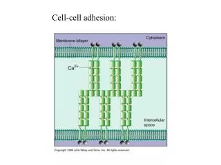
cell
Single Cell Analysis with an Integrated Electrophoretic/ Electrochemical Chip. Ching-Yu CHANG 1, 2 , Tatsuya MURATA 2 , Yasufumi TAKAHASHI 2 , Ryota KUNIKATA 2 , Hitoshi SHIKU 2 , Hsien-Chang CHANG 1 , Tomokazu MATSUE 2 *.

cell
E N D
Presentation Transcript
Single Cell Analysis with an Integrated Electrophoretic/ Electrochemical Chip Ching-Yu CHANG1, 2, Tatsuya MURATA2, Yasufumi TAKAHASHI2, Ryota KUNIKATA2, Hitoshi SHIKU2, Hsien-Chang CHANG1, Tomokazu MATSUE2* 1-Institute of Biomedical Engineering, National Cheng Kung University, Tainan 701, Taiwan 2-Graduate School of Environmental Studies, Tohoku University, Sendai 980-8579, Japan conductive substrate redox recycling S catalyze Pred Poxi secretion cell electrode Oxidation current
Electrochemical Single Cell Measurement Interference: + + SNR: - Signal level: - Interference: + + SNR: - Signal level: - Interference: - - SNR: + + Signal level: + + Interference : - SNR: + Signal level: +
Micro Well Structure 100 mm 30 mm 5 mm 15mm LFVA (low flow velocity area) 25 mm
Active vs. Passive Single Cell Trap How to take advantages of active and passive traps?
Chip Design 30 mm SU- 8 ITO 25 mm Pt 30 mm + - +2.0 V tape SU-8 electrode Trapping solution: 0.2 M sucrose Trapping voltage: 2.0 V vs. ITO
Cell Manipulation Top View Electrophoretic trapping - + - 2.0 V Hydraulic flush Electrophoretic repelling + - + 2.0 V
Steady-state Currents for Microelectrode i Ilim E disc electrode parameters n: transferring electron / molecule F: Faraday constant D: diffusion constant C: substance conc. r: electrode radius L: recessed depth recessed disc electrode Analyst, 2004 (129) 1157-65
Model for Recessed UME on a Conductive Substrate l r d conductive substrate redox normalized parameter H=l/r, L=d/r UME Chip Electrode Anal. Chem., 2007 (79) 5809-16
Micro Well Electrode Validation < 30 mm ~ 12.9 mm L=23 mm 5.5 nA scan rate: 10 mV/sec 2E configuration, Ag/AgCl as RE+CE 5 mM K3Fe(CN)6 / 0.1 M KCl scan direction: 0.6 0 0.6 V 7.8 nA I T = 1.07 (theoretical) I T =7.8/5.5= 1.42
SEAP p-aminophenol (PAP) p-aminophenylphosphate (PAPP) 0.3 V vs. Ag/AgCl + 2H+ +2 e- 0.1 V vs. Ag/AgCl p-iminoqulnone (IQ) PAP NH2 HO NH2 O Measurement of Secreted Alkaline Phosphatase ITO electrode PAPP/HEPES diffusion PAP IQ diffusion PAPP PAP PAP IQ SEAP recombinant HeLa e- Pt electrode SEAP: secreted alkaline phosphatase
Redox Recycling on ITO Electrode detection voltage Dot line RE+CE ITO PAP WE Solid line RE+CE WE PAPP Measuring condition PAP 4.7 mM /HEPES (line a &b) PAPP 4.7 mM /HEPES (line c &d) HEPES buffer: HEPES 20 mM, NaCl 153 mM, KCl 5 mM, glucose 5 mM, pH 9.5 scan rate: 20 mV/sec scan direction: 0 0.6 0 V
ALP-Bead Preparation + 0.3 V vs. Ag/AgCl n=3 UME RE+CE wash with HEPES, suspend in 2.35 mMPAPP particle descending latex bead: 10 mm HEPES: pH 9.5 ALP (0.6U/mL) / HEPES overnight incubation
Single ALP-Bead Measurement iPAP depletion PAPP PAP bare bead ALP bead
Real-time SEAP Secretion Monitoring SEAP Cell Micro well PAP calibration curve
Conclusion • Cell can be trapped and repelled by electrophoretic force. • Micro well structure can provide a LFVA to stabilize the trapped cell during solution change. • ITO electrode provide a conductive surface for redox recycling and then enhances the response current. • The real-time non-continuous SEAP secretion was observed by this device. Thanks for your attention …
Entrapment and measurement of a biologically functionalized microbead with a microwell electrodeChing-Yu Chang, Yasufumi Takahashi, Tatsuya Murata, Hitoshi Shiku, Hsien-Chang Chang* and Tomokazu Matsue* Lab Chip, 2009, 9, 1185–92
1 M H2SO4 1 M NaOH
不同電位電析Pd粒子於GC電極上的型態 電極 1 電極 2 電極 3 電極 1 電極 2 電極 3 電極 4
電位階昇法:不同電透析條件下Pd(GOD)/GC電極於PBS(pH 7.4)中的循環伏安圖
Amino Acid Structures http://www.cem.msu.edu/~cem252/sp97/ch24/ch24aa.html
pKa Values of Amino Acid http://www.cem.msu.edu/~cem252/sp97/ch24/ch24aa.html