Download
1 / 16
160 likes | 320 Vues
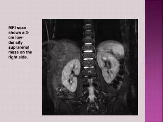
MRI scan shows a 3-cm low-density suprarenal mass on the right side. Pheochromocytoma. Myra Lalas Pitt. definition. Catecholamine-secreting tumors that arise from chromaffin cells located in the adrenal medulla and sympathetic chain.

E N D
MRI scan shows a 3-cm low-density suprarenal mass on the right side.
Pheochromocytoma Myra Lalas Pitt
definition • Catecholamine-secreting tumors that arise from chromaffin cells located in the adrenal medulla and sympathetic chain. • Associated with neurofibromatosis type 1, familial paraganglioma, von Hippel-Lindau disease, tuberous sclerosis, Sturge-Weber syndrome, and multiple endocrine neoplasia (MEN 2A, 2B).
von Hippel-Lindau disease • Inherited, AD syndrome manifested by a variety of benign and malignant tumors
MEN 2A Heritable predisposition to medullary thyroid cancer, pheochromocytoma, and primary parathyroid hyperplasia. • MEN 2B Predisposition to medullary thyroid cancer, pheochromocytoma, mucosal neuromas Constipation, megacolon, developmental abnormalities, Marfanoid habitus, myelinated corneal nerves
Signs and symptoms • Sweating • Episodic headaches • Tachycardia • Hypertension • Pallor • Nausea/vomiting • Weakness • Abdominal pain • Anxiety • Chest pain Classic triad
Differentials • Glomerulonephritis • Renal vein thrombosis • Coarctation of the aorta • Hyperthyroidism • Cushing Syndrome • Meds (TCA’s) • Cocaine use • Neuroblastoma • Acute intermittent porphyria • Idiopathic Intracranial Hypertension
Diagnostic studies • Plasma free metanephrine (100% sensitive; 96.7% specific with negative predictive value 100%) • 24-h urine for epinephrine, norepinephrine, dopamine, metanephrines, homovanillic acid, vanillylmandelic acid, creatinine (sensitivity of 87.5% and a specificity of 99.7%)
Adrenal CT • MRI- preferred due to the high sensitivity for adrenal tumors and better ability to localize extra-adrenal pheochromocytomas • Chem 10 • 123I-metaiodobenzylguanidine (MIBG) scan to screen the body- used to locate and confirm pheochromocytoma and rule out metastatic disease • PET scan.
Large left adrenal mass and a lesion in the right hepatic lobe
Treatment • Laparoscopic surgery is now the technique of first choice for resection adrenal and extra-adrenal pheochromocytomas. • All patients receive appropriate preoperative medical management to block the effects of released catecholamines.
After adequate alpha-adrenergic blockade has been ensured (PHENOXYBENZAMINE), beta-adrenergic blockade is started, which typically occurs 2-3 days preoperatively. • Beta-adrenergic blockade should never be started first. • Blockade of vasodilatory peripheral beta-adrenergic receptors with unopposed alpha-adrenergic receptor stimulation can lead to a further elevation in blood pressure.
QUESTION • A 14-yo female undergoing evaluation for persistent headaches and vomiting complains of double vision with lateral gaze. On PE, she is alert, but fundoscopic exam reveals blurring of the disk margin and absence of venous pulsations. A head MRI is normal. LP reveals an elevated opening pressure, with normal glucose, protein, and cells.
What is the most common long term complication of this girl’s condition? • Progressive hemiparesis • Generalized seizures • Urinary incontinence • Visual loss • Facial nerve palsy
ANSWER: D • Pseudotumor cerebri: Optic nerve atrophy and visual loss are significant complications.
References Chernausek SD, Stratakis CA. Chapter 537. Endocrine Neoplasia Syndromes. In: Rudolph CD, Rudolph A, Lister GE, First L, Gershon A, eds. Rudolph's Pediatrics. 22nd ed. New York: McGraw-Hill; 2011. http://www.accesspediatrics.com/content/7054216. Accessed February 2, 2012. Lowry AW, Bhakta KY, Nag PK. Chapter 14. Endocrinology. In: Lowry AW, Bhakta KY, Nag PK, eds. Texas Children's Hospital Handbook of Pediatrics and Neonatology. New York: McGraw-Hill; 2011. http://www.accesspediatrics.com/content/7436745. Accessed February 2, 2012. Pediatrics in Review Vol. 32 No. 6 June 1, 2011 pp. 257 -263 (doi: 10.1542/pir.32-6-257) www.uptodate.com