Advances in Electron Microscopy: Resolution and Imaging Techniques
Electron microscopes, developed in the 1950s, revolutionized imaging with their ability to produce two-dimensional images using accelerated electrons at a voltage of 105V. These electrons have a wavelength of about 0.004nm, enabling practical resolutions of 1 to 5nm, significantly surpassing visible light's resolution. Notably, the Scanning Electron Microscope (SEM), commercialized in 1965, offers resolutions down to 1nm laterally and 0.1nm vertically, utilizing quantum tunneling techniques. The Bohr model also plays a crucial role in understanding atomic spectra and photon energy transitions.

Advances in Electron Microscopy: Resolution and Imaging Techniques
E N D
Presentation Transcript
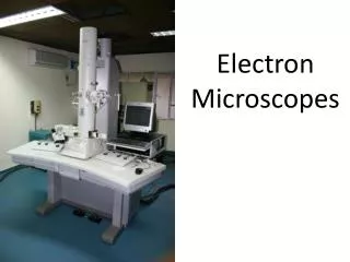
Electron Microscopes 27-7
Developed in the 1950s • Two Dimensional Image • Electrons are accelerated by a Voltage of 105V • These Electrons have a wavelength of about .004nm • Practical resolution is .1 to .5nm • This is still 103 better than visible light • 400-800nm Transmission Electron Microscope
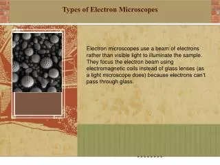
Resolutions to 1nm • Produce 3D image • 1st commercial product 1965 Scanning Electron Microscope
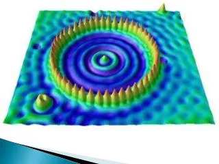
Developed in the 1980s • .1nm resolution laterally • .01nm resolution vertically • Uses Quantum Tunneling 30-12 Scanning Tunneling EM
Atomic Spectra 27-11
Bohr Model relates the jump down between excited states of the atom to the energy of a photon • Each line of the spectrum is a photon step • The larger the step, the more energy the photon • Therefore the shorter the wavelength • And higher frequency Bohr Model
Rydberg Constant • R = 1.0974 x 10⁷m-1 • Balmer Series • Lyman Series • Paschen Series Hydrogen Series