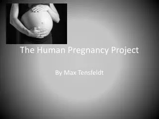
Pregnancy and Human Development
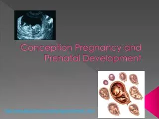
28. P A R T B. Pregnancy and Human Development. Organogenesis. Gastrulation sets the stage for organogenesis, the formation of body organs By the 8 th week all organ systems are recognizable. Specialization of Ectoderm.


Pregnancy and Human Development
E N D
Presentation Transcript
28 P A R T B Pregnancy and Human Development
Organogenesis • Gastrulation sets the stage for organogenesis, the formation of body organs • By the 8th week all organ systems are recognizable
Specialization of Ectoderm • Neurulation – the first event of organogenesis gives rise to the brain and spinal cord • Ectoderm over the notochord thickens, forming the neural plate • The neural plate folds inward as a neural groove with prominent neural folds
Specialization of Ectoderm • By the 22nd day, neural folds fuse into a neural tube, which pinches off into the body • The anterior end becomes the brain; the rest becomes the spinal cord • Associated neural crest cells give rise to cranial, spinal, and sympathetic ganglia
Specialization of Ectoderm: Neuralization Figure 28.9a, b
Specialization of Ectoderm: Neuralization Figure 28.9c, d
Specialization of Endoderm • Embryonic folding begins with lateral folds • Next, head and tail folds appear • An endoderm tube forms the epithelial lining of the GI tract • Organs of the GI tract become apparent, and oral and anal openings perforate • Endoderm forms epithelium linings of the hollow organs of the digestive and respiratory tracts
Folding of the Embryonic Body Figure 28.10a–d
Endodermal Differentiation Figure 28.11
Specialization of the Mesoderm • First evidence is the appearance of the notochord • Three mesoderm aggregates appear lateral to the notochord • Somites, intermediate mesoderm, and double sheets of lateral mesoderm
Specialization of the Mesoderm • The 40 pairs of somites have three functional parts: • Sclerotome – produce the vertebrae and ribs • Dermatome – help form the dermis of the skin on the dorsal part of the body • Myotome – form the skeletal muscles of the neck, trunk, and limbs
Specialization of the Mesoderm • Intermediate mesoderm forms the gonads and the kidneys • Lateral mesoderm consists of somatic and splanchnic mesoderm
Specialization of the Mesoderm • Somatic mesoderm forms the: • Dermis of the skin in the ventral region • Parietal serosa of the ventral body cavity • Bones, ligaments, and dermis of the limbs • Splanchnic mesoderm forms: • The heart and blood vessels • Most connective tissues of the body
Specialization of the Mesoderm Figure 28.12
Development of Fetal Circulation • By the end of the 3rd week: • The embryo has a system of paired vessels • The vessels forming the heart have fused
Development of Fetal Circulation • Unique vascular modifications seen in prenatal development include umbilical arteries and veins, and three vascular shunts (occluded at birth) • Ductus venosus – venous shunt that bypasses the liver • Foramen ovale – opening in the interatrial septa to bypass pulmonary circulation • Ductus arteriosus – transfers blood from the right ventricle to the aorta
Circulation in Fetus and Newborn Figure 28.13
Effects of Pregnancy: Anatomical Changes • Chadwick’s sign – the vagina develops a purplish hue • Breasts enlarge and their areolae darken • The uterus expands, occupying most of the abdominal cavity
Effects of Pregnancy: Anatomical Changes • Lordosis is common due to the change of the body’s center of gravity • Relaxin causes pelvic ligaments and the pubic symphysis to relax • Typical weight gain is about 29 pounds
Relative Uterus Size During Pregnancy Figure 28.15
Effects of Pregnancy: Metabolic Changes • The placenta secretes human placental lactogen (hPL), also called human chorionic somatomammotropin (hCS), which stimulates the maturation of the breasts • hPL promotes growth of the fetus and exerts a maternal glucose-sparing effect • Human chorionic thyrotropin (hCT) increases maternal metabolism • Parathyroid hormone levels are high, ensuring a positive calcium balance
Effects of Pregnancy: Physiological Changes • GI tract – morning sickness occurs due to elevated levels of estrogen and progesterone • Urinary system – urine production increases to handle the additional fetal wastes • Respiratory system – edematous and nasal congestion may occur • Dyspnea (difficult breathing) may develop late in pregnancy
Effects of Pregnancy: Physiological Changes • Cardiovascular system – blood volume increases 25-40% • Venous pressure from lower limbs is impaired, resulting in varicose veins
Parturition: Initiation of Labor • Estrogen reaches a peak during the last weeks of pregnancy causing myometrial weakness and irritability • Weak Braxton Hicks contractions may take place • As birth nears, oxytocin and prostaglandins cause uterine contractions • Emotional and physical stress: • Activates the hypothalamus • Sets up a positive feedback mechanism, releasing more oxytocin
Parturition: Initiation of Labor Figure 28.16
Stages of Labor: Dilation Stage • From the onset of labor until the cervix is fully dilated (10 cm) • Initial contractions are 15–30 minutes apart and 10–30 seconds in duration • The cervix effaces and dilates • The amnion ruptures, releasing amniotic fluid (breaking of the water) • Engagement occurs as the infant’s head enters the true pelvis
Stages of Labor: Dilation Stage Figure 28.17a, b
Stages of Labor: Expulsion Stage • From full dilation to delivery of the infant • Strong contractions occur every 2–3 minutes and last about 1 minute • The urge to push increases in labor without local anesthesia • Crowning occurs when the largest dimension of the head is distending the vulva
Stages of Labor: Expulsion Stage Figure 28.17c
Stages of Labor: Expulsion Stage • The delivery of the placenta is accomplished within 30 minutes of birth • Afterbirth – the placenta and its attached fetal membranes • All placenta fragments must be removed to prevent postpartum bleeding
Stages of Labor: Expulsion Stage Figure 28.17d
Extrauterine Life • At 1-5 minutes after birth, the infant’s physical status is assessed based on five signs: heart rate, respiration, color, muscle tone, and reflexes • Each observation is given a score of 0 to 2 • Apgar score – the total score of the above assessments • 8-10 indicates a healthy baby • Lower scores reveal problems
First Breath • Once carbon dioxide is no longer removed by the placenta, central acidosis occurs • This excites the respiratory centers to trigger the first inspiration • This requires tremendous effort – airways are tiny and the lungs are collapsed • Once the lungs inflate, surfactant in alveolar fluid helps reduce surface tension
Occlusion of Fetal Blood Vessels • Umbilical arteries and vein constrict and become fibrosed • Fates of fetal vessels • Proximal umbilical arteries become superior vesical arteries and distal parts become the medial umbilical ligaments • The umbilical vein becomes the ligamentum teres • The ductus venosus becomes the ligamentum venosum • The foramen ovale becomes the fossa ovalis • The ductus arteriosus becomes the ligamentum arteriosum
Transitional Period • Unstable period lasting 6-8 hours after birth • The first 30 minutes the baby is alert and active • Heart rate increases (120-160 beats/min.) • Respiration is rapid and irregular • Temperature falls
Transitional Period • Activity then diminishes and the infant sleeps about three hours • A second active stage follows in which the baby regurgitates mucus and debris • After this, the infant sleeps, with waking periods occurring every 3-4 hours
Lactation • The production of milk by the mammary glands • Estrogens, progesterone, and lactogen stimulate the hypothalamus to release prolactin-releasing hormone (PRH) • The anterior pituitary responds by releasing prolactin
Lactation • Colostrum • Solution rich in vitamin A, protein, minerals, and IgA antibodies • Is released the first 2–3 days • Is followed by true milk production
Lactation and Milk Let-down Reflex • After birth, milk production is stimulated by the sucking infant Figure 28.18
Breast Milk • Advantages of breast milk for the infant • Fats and iron are better absorbed • Its amino acids are metabolized more efficiently than those of cow’s milk • Beneficial chemicals are present – IgA, other immunoglobulins, complement, lysozyme, interferon, and lactoperoxidase • Interleukins and prostaglandins are present, which prevent overzealous inflammatory responses • Its natural laxatives help cleanse the bowels of meconium