Download
1 / 13
150 likes | 407 Vues
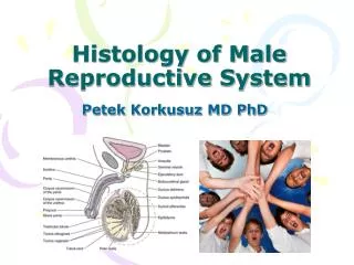
Histology of the Male Genital System. The Male Genital System. The male genital system consists of: Primary sex organ : two testes. Accessory sex organs : accessory glands and excretory ducts. Accessory glands: seminal vesicle, prostate, bulbo-urethral glands.

E N D
The Male Genital System • The male genital system consists of: • Primary sex organ: two testes. • Accessory sex organs: accessory glands and excretory ducts. • Accessory glands: seminal vesicle, prostate, bulbo-urethral glands. • Excretory ducts: tubuli recti, rete testis, ductuli efferentes, epididymis, vas deferens, ampulla of vas deferens, ejaculatory duct and urethra.
The Prostate • It is the largest accessory genital gland in males. • It is a compound tubulo-alveolar gland which is pierced by the prostatic urethra and ejaculatory ducts. • The ducts of the gland opens open into the prostatic urethra. • It has connective tissue stroma and parenchyma.
The Stroma • The slender connective tissue capsule is composed of a richly vascularized, dense irregular collagenous connective tissue, interspersed with smooth muscles fibers. • The stroma of the gland is derived from the capsule, so, it is rich in smooth muscle fibers in addition to the normal connective tissue.
The glandular parenchyma • The prostatic acini with irregular cavities varying in shape and size and epithelial lining which may be simple columnar or pseudostratified columnar depending upon the activity of the gland. • The prostate is strongly positive for acid phosphatase enzyme which is taken as indicator for the gland activity. • The prostatic acini have characteristic prostatic concretion which are acidophilic concentric lamellae of accumulated secretions that may be calcified in old age.
Functions of the prostate • It gives the fluid medium of the semen which contains: • Hydrolytic enzymes which include acid phosphatase and zinc which has two functions: • Energy preserving effect on spermatozoa. • Protective effect against phagocytic cells in the female genital tract • But zinc content inside the prostate makes it liable to chronic bacterial inflammation due to inhibition of its own phagocytic cells.
Parts of the prostate • Prostate proper: • Central zone. • Peripheral zone. • Transitional zone. • Periurethral tissue. • Prostatic utricle. • The peripheral zone is the main site of the prostatic carcinoma. • The transitional zone is the main site for benign prostatic hypertrophy which may compress the urethra causing urine retention.
Prostate proper: Both central and peripheral zones lie posterior to the urethra and anterior to the ejaculatory ducts. The central zone is related to the neck of the urinary bladder. • Transitional zone: it lies anterior to the peripheral part and resembles it except that its stroma is compact and fibrous. • Periurethral tissue: small glandular diverticula of the upper part of the prostatic urethra. • Prostatic utricle: a small pouch located in the seminal colliculus above the entrance of the ejaculatory duct. It is the remnant of fused mullarian ducts.
Erectile tissue of the Penis • The erectile tissue of the three corpora of the penis consists of irregular cavities lined by endothelium and separated by connective tissue trabeculae containing elastic fibers and smooth muscle fibers and sensory nerve endings. The endothelial cavities are continuous with the arterial supply and venous drainage. • Erection is done by loss of muscle tone in the arterial walls that allow more blood to fill the cavities of the erectile tissue, resulting in expansion of these cavities which compress the veins and prevent its drainage and increase the size of the spongy penis resulting in erection.