Download
1 / 22
360 likes | 1.35k Vues
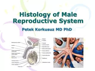
Histology of male reproductive system. Functions of the male reproductive system:

E N D
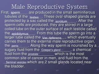
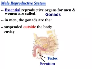
Functions of the male reproductive system: 1. Ensuring the development of germ cells2. Copulation is elimination of germ cells3. Biosynthesis of male sex hormonesGroups of male reproductive system:1) gonads - testes (1)2) The bodies of the deposit of extratesticular (appendage (2), the vas deferens (3), ejaculatory canal)3) additional sex organs are the seminal vesicles (4), prostate (5), penis (penis, 7).
The development of the male reproductive system.I.Indifferent stage is for the 4th week. embryogenesis on the inner side of the primary kidney coelomic epithelium formed genital ridges. They migrate from the yolk sac gonoblast. The epithelium of genital ridges multiplies and grows into the primary kidney cortex is formed sex cords. At the same time from the flow splits mezonephral paramezonephral duct.
Stage II of differentiation of the gonads of male type - with 6 weeks of embryogenesis of sex cords formed convoluted, straight tubules of the testis and tubules of the testis network.Mezonephral of the seminiferous duct develop ways and prostate gland.Paramezonephral duct undergoes regression.
TesticlesFunctions:1. Generative: development of spermatozoa.2. Endocrine: development of male sex hormones.The structure of the testisTestis is lobed parenchymal organ.Shell eggs: serous, tunica, vascular. From the mediastinal connective tissue radiating trabeculae, which divide the egg into slices. Strom presented LFCT.
Testicular parenchyma was formed crimped, straight seminiferous tubules and tubule network. The number of lobules in the testis ≈ 200. In the clove - 1-4 convoluted tubule, L = 80 cm total length of the convoluted tubules 160-640m. Convoluted tubules pass into lines, which merge to form the rete testis.
Structural and functional unit of the testis is twisted spermatic tubule.From the outside it is covered by your own shell:The inner fibrous layer is the network of collagen fibers.Myoidal layer myoidal cells, reducing the convoluted tubule.The outer fibrous layer: basal cell membrane mioidnyh + network of collagen fibers with fibrocytes.
Epiteliospermatogenic layer. Composed sustentocytes or Sertoli cells and developing germ cells, of which the basement membrane in contact only spermatogonia.
Sertoli cellshave a triangular shape, the base lies on the basal membrane. Sharp peaks of Sertoli cells with spikes appear in the lumen of convoluted tubule. Outgrowths of adjacent Sertoli cells are connected with each other desmosomes. The lumen of the tubule is divided into 2 floor. On the ground floor are the spermatogonia, the rest of the developing male germ cells are in the second floor.
Function of Sertoli cells1. Trophism of developing germ cells2. Reference3. Hormonal and secretory (synthesis of inhibin, activin)4. Participation in Education blood-testis barrier (BTB)5. The transport functionSertoli cells are resistant to damaging factors, more than spermatogenic cells. After exposure to ionizing radiation convoluted tubule seed can contain only Sertoli cells (syndrome "Only Sertoli cells").
BTB functions: 1. Prevention of autoimmune reactions.2. Prevention or reduction of income gametes damaging chemical and biological agents.3. Transport of nutrients and regulatory substances.4. Microenvironment for germ cells of different maturity.GTB can be damaged by trauma testis, mumps, in some cases there is an autoimmune orchitis in which germ cells die. BTB penetrate through alcohol, nicotine, which have deleterious effects on spermatogenesis.
Spermatogenesis includes 4 phases:1. Reproduction 2. Growth3. Maturation 4. formationSpermatogonia are powered from the capillaries through the BTB.Spermatocytes I order separate from the basement membrane and move in 2 floor convoluted tubule.Spermatocytes of the I form spermatocytes II and spermatids, which are transformed into sperm. All of these cells lie in the 2ndfloor of the tubule and are powered by Sertoli cells. Along the length of seminiferous tubules spermatogenesis is simultaneously.
Spermatogenesis takes ≈ 75 days, the longest phase formation takes place, or spermatogenesis. Spermatogenesis is sensitive to the action of harmful factors: stress, smoking, alcohol, ionizing radiation. Spermatogenesis also inhibited when exposed to elevated temperatures, for example, when making a man of common hot tubs, cryptorchidism (abdominal temperature ≈ 3-4 degrees higher than in the scrotum). Determine which of the histological sections showed inhibition of testicular spermatogenesis in cryptorchidism.
Endocrine testicular functionIn the testes are formed male sex hormones that stimulate sperm production, development of secondary sexual characteristics, muscle growth and shape the sexual behavior of men (libido). They are produced in the interstitial Leydig endocrinocytes. Number of Leydig cells to puberty are 700mln, Every 10 years is reduced by 80mln due to apoptosis.
Deferent way’sfunctions:Deposit, trophism, air conditioning (making the state of readiness for fertilization), sperm.Providing a massive release of semen during intercourse.The secretory.Endocrine.By the seminiferous pathways include direct canals, canals network, delivering tubules head appendages, the channel appendage, and ejaculatory duct vas, urethra.
Straight tubules lined by a single layer of cuboidal or columnar epithelium. On the apical surface of epithelial cells have microvilli and solitary cilia. Under the epithelium lies with the individual's own record smooth myocytes.Network testis is lined with flat or cuboidal epithelium. Cells have different heights and on the apical surface are single cilium. Outside the epithelium is its own record with a single smooth myocytes.
Efferent ducts are in the head of the epididymis. Have three shells: the mucosa, muscle and adventitial. The epithelium contains two types of cells: ciliated and secretory secreting holocrine in type. These cells have different heights, lie groups, resulting in the lumen of the tubule is uneven. The epithelium rests on a thin plate of its own mucous membranes. Muscle membrane is formed by several layers of smooth myocytes, and adventitia isLFCT.
The channel is an appendage of the body of the epididymis. The mucous membrane is represented seriate epithelium of prismatic cells with stereocilia and intercalary (basal) cells and the lamina propria. Shaped like tongues of flame through the speakers of stereocilia ("flaming epithelium"). Muscle and adventitia formed smooth muscle tissue and LFCT. The epithelium of the epididymis produces a fluid channel, diluted sperm factor and longitudinal motility.
The vas deferens is the mucosa, muscle and adventitia. Epithelium-row (ciliated and intercalated cells). Lamina propriaisLFCT. The muscular sheath consists of inner and outer longitudinal and middle circular layers. Adventitia is represented byLFCT.Between the confluence of the vas deferens and seminal vesicles is the beginning of the urethra ejaculatory canal, built in the same way as the vas deferens, but it is thinner muscle membrane.
Prostate.Glandular and muscular organ that surrounds the urethra. It is composed of 30-50 tubular - alveolar glands. In the end parts of the single row, double row sometimes epithelium containing secretory and intercalated cells. In the pseudostratified epithelium of the excretory ducts, and at the confluence of the urethra is transitional. 50% of the prostate is smooth muscle tissue, which lie between the cells of elastic fibers. Smooth myocytes surround the terminal units, ducts, and ensure the release of secret during coitus.Functions is to develop a secret, diluting the semen containing sperm activating and nutritional factors
Agechangesof malereproductive system. Testicle of Newbornand children under 4 yearsseminiferous tubuleshave the form of continuouscell cordsconsisting of Spermatogoniaand sustentocytes.From 4 to 10 years (during growth) convoluted tubules more twists, they appear light. From 5 to 6 years, there dividing spermatogonia. To 10.9 years appear first spermatocyte I order. From 10 to 12 years - the period of development, there is growth of the seminiferous tubules by increasing the number and size of cells. Revealed spermatocytes I and II of the order. By 12-16 years (the period of the final formation of spermatogenesis) completes the development of the testis.Testicular involution begins after age 50 and older in terms of the seminiferous tubules spermatogenesis is preserved, the structure of the tubules may be close to normal.
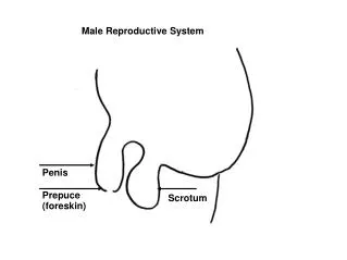
Penis. Outside, covered with a thin skin. In the penis there are three elongated erectile (cavernous) of the body is two on each side, joined in the middle part, and under them is the corpus cavernosum urethra. Outside they are covered with tunica from DFCT, inside depart incomplete trabeculae. Between the erectile tissue is connective tissue. Blood to the cavernous bodies replenished by bringing arteries and efferent veins flowing in, which lie under the tunica, then pierce it and go lightly.An erection is caused by an accumulation of blood in vascular spaces as a result of the predominance of inflows over outflows and squeezing the efferent venules.Cavernosum urethra consists of DFCT, which has a lot of convoluted veins. Head of the penis is an extension of the corpus cavernosum urethra. Fold of skin covering the head is the foreskin.