Download
1 / 24
270 likes | 604 Vues
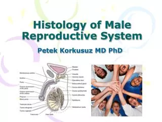
Histology of Male Reproductive System. Lecture Objectives. Describe the histological features of the male reproductive system. Male Reproductive System. The male structures of reproduction include the: testes , a system of ducts ductus epididymis ductus deferens ejaculatory duct

E N D
LectureObjectives • Describe the histological features of the male reproductive system
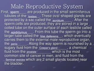
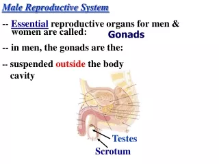
Male ReproductiveSystem • The male structures of reproduction includethe: • testes, • a system ofducts • ductus epididymis • ductus deferens • ejaculatoryduct • urethra • accessory sexglands • seminalvesicles • prostategland • bulbourethralglands • supportingstructures • penis
Testis: GeneralOrganization • Tunica vaginalis – extension ofperitoneum • Tunica albuginea – dense CT layer • Mediastinum – projectionof the tunicaalbuginea • Lobules – formed fromsepta from the tunicaalbuginea
SeminiferousTubules • General structure – 200-500 m inlength • Tunica propria – lamina propria surrounding the seminiferoustubules • Myoid cells – 3-5 layers thick, synthesize collagen, contractions help move sperm through thetubules • Basallamina • Seminiferous epithelium – complex stratified epithelium composed of spermatogenic cells and sertolicells
Spermatogeniccells • Type A spermatogonia – ovoid nuclei, can be eithertype A dark (stem cells) or type A pale (committed tobecome sperm) • Type B spermatogonia – formed by mitotic division of type A pale cells, connected to each other by cytoplasmicbridges • Primary spermatocytes – result from the mitotic division of type B spermatogonia, large nuclei with dark staining condensedchromatin • Secondary spermatocytes – formed from the first meiotic division of primaryspermatocytes • Spermatids – formed following the second meiotic division • Spermatozoa – formed followingspermiogenesis
Sertoli (Supporting)Cells • Structure • Long cytoplasmic processes extending between spermatogenic cells • Contain inclusion bodies ofCharcot-Böttcher • Functions • Blood-testis barrier – formed from unique tight junctions, separates sperm from immunesystem • Hormones – release inhibin, androgen bindingproteins • Respond to FSH andtestosterone • Support – support and nourish spermatocytes, therefore, also known as supporting or sustentacularcells • Other characteristics – do notreplicate
LeydigCells • Structure • large, polygonal,acidophilic • Contain lipid droplets & Reinkecrystals • Location – interstitium of seminiferoustubules • Function – secrete testosterone in response toLH • Do notreplicate • Produce testosterone during early fetal life but are inactive untilpuberty
Spermatogenesis • Note: during spermatogenesis, the developing sperm are connected to each other by cytoplasmic bridges. This ensures synchronous development of eachclone • Spermatogonial phase(mitosis) • Type A darkm→itosisType A palemi→tosisType A palem→itosisType Bm→itosis primaryspermatocytes • Spermatocyte phase(meiosis) • Meiosis I – primary spermatocyte forms two secondary spermatocytes (separation ofchromosomes) • Meiosis II – secondary spermatocyte forms two spermatids containing haploid DNA (separation ofchromatids)
Spermiogenesis • there are four phases: • Golgiphase • Proacrosomal granules – contain glycoproteins (enzymes); granules fuse to form the acrosomalvesicle • Acrosomal vesicle – located near the nuclear membrane; its location determines the anterior pole of the developingsperm • Axonal complex – migration of centrioles to the posterior pole to initiate synthesis of axonemal complex of spermtail • Cap phase • Acrosomal cap – acrosomal vesicle condenses and covers the anterior half ofnucleus • Acrosome phase • Reorientation of spermatid with head pointed down into Sertolicell • Lengthening offlagellum • Manchette – organization of cytoplasmicmicrotubules • Centrioles migrate back to nucleus to initiate formation of the neck region • Maturation phase • Residual body – excess cytoplasm is pinched off and phagocytized by Sertolicells
Structureofa Mature Sperm • Head – nucleus & acrosomalcap • Neck – centriole, excess nuclear envelop, striated columns • Tail • Middle piece – 9 outer doublets, central doublet of MTs, 9 outer dense fibers, mitochonderialsheath • Principle piece – outer dense fibers gradually decrease in number and are surrounded by a fibroussheath • Tail end piece – microtubules, no densefibers
IntratesticularDucts • Straight Tubules (Tubulirecti) • Location – immediately after seminiferoustubule • Epithelial lining – Sertoli cells only at junction with seminiferous tubules becoming simple cuboidal near retetestis • Function – transport ofsperm • Rete Testis • Location – interconnecting within CT ofmediastinum • Epithelial lining – simple cuboidal to lowcolumnar • Function –transport • EfferentDuctules • Location – rete testis connects to 20 efferentductules • Epithelial lining – pseudostratified columnar (sawtooth appearancedue to presence of tall ciliated columnar cells used for moving sperm and low columnar cells with microvilli used for fluid absorption; basal cells are alsopresent) • Muscle layer – first appearance of a layer of smooth muscle; elastic fibers are interspersed among the musclecells • Function – transport ofsperm
Ductusepididymus • Efferent ductules coalesce to form a single ductus epididymis Structure– highly coiled; 4-6 mlong • Divisions – head, body,tail • Tail is the principle reservoir of maturesperm • Epithelium – pseudostratified columnar withstereocilia • Muscular layer – graduallythickens • the tail has three layers: inner and outer longitudinal and middle circular layer. • The muscle of the head and body move sperm by peristaltic contraction; sperm is ejected during ejaculation by muscle of thetail • Function • Sperm reservoir (tailregion) • Fluid absorption and phagocytosis of remaining residual bodies and damaged sperm • Secretion • Further development of sperm – addition of surface glycosides to inhibit sperm binding toegg • Ejection of sperm duringejaculation
Vasdeferens • Structure • Epithelium – pseudostratified columnar withmicrovilli • Three muscle layers – inner & outer longitudinal, middlecircular • Ampulla – enlargement of the distal end where it is joined by the duct of the seminalvesicle • Functions – secretory;transport Ejaculatoryducts • Structure – continuation of the vas deferens through the prostategland • Epithelium - pseudostratified columnar withmicrovilli • Other features– • NO muscle layers except fibromuscular tissue of theprostate • Empties into the prostaticurethra • Functions – secretory;transport
vasdeferens Epididymis
SeminalVesicles • Structure • One primary lumen; numerous primary and secondary folds that increase surface area • Pseudostratified columnarepithelium • Inner circular and outer longitudinal muscle layers which contracts during emission adding stored secretions to seminalfluid • Function • produces and secretes 60% of the volume ofsemen • secretion is viscous, whitish-yellow containing fructose, simple sugars, ascorbic acid, amino acids and prostaglandins. • Secretion requirestestosterone
ProstateGland • Structure– • Consists of 30-50 tubuloalveolarglands • lined with pseudostratified or simple columnarepithelium. • The glandular components are arranged in threelayers: • Mucosal layer (central zone) – empty directly in theurethra • Submucosal layer (transition zone) – glands empty first into the prostatic sinus and then into theurethra • Peripheral layer (peripheral zone) – the main prostatic glands, empty into prostatic sinuses • Prostatic concretions (corpora amylacea) – precipitated secretory material that may be calcified, found in alveoli particularly in oldermen • Capsule – fibroelastic CT and smooth muscle, contracts duringemission • Function– produces 30% of semenvolume. • Secretion is milky fluid containing citric acid, cholesterol, acid phosphatase, fibrinolysin, andelectrolytes • Benign prostatic hypertrophy (BPH) &cancer • BPH normally occurs in older men in the mucosal and submucosalglands • malignancies occur in the epithelium of the main peripheralglands
BulbourethralGlands • Structure – tubuloalveolar, mucus secretory glands lined with simple columnarepithelium • – Surrounded by intermixed smooth & skeletalmuscle • Function – secretes preseminal fluid that lubricates the penile urethra prior toejaculation • Glands of Littre (urethralglands) • Structure – predominantly around penileurethra • Function – mucus secretion protectsepithelium from urine
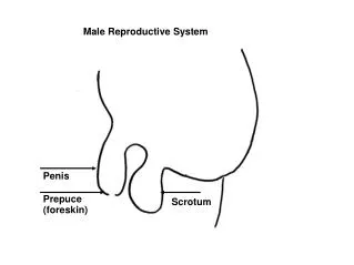
Penis • Structure • Erectile tissue – three cavernous bodies surrounded by loose elastic connective tissue with an outer covering of thinskin • skin extends over the glans penis (prepuce or foreskin) unless removed by circumcision. • A thick fibrous sheath called the tunica alboginea surrounds each cavernousbody. • The cavernous bodies consist of irregular vascular spaces separated by trabeculae of dense fibroelastictissue. • The 3 bodiesare: • Corpora cavernosa – two dorsal erectile tissues of thepenis • Corpus spongiosum (corpus cavernosum urethrae) – ventral erectile tissue, surroundsurethra
Erection • Results from parasympathetic postganglionic efferent impulses which cause penile arteries to dilate, more blood enters cavernous spaces, cavernous body distends compressing veins, engorgement results inerection • arterial dilation also mediated by nitric oxide produced by endothelial cells • Note: Viagra enhances the effects of nitric oxide resulting in vasodilation • Emission • Results from adrenergic (sympathetic)stimulation • stimulation causes movement of sperm from the tail of the epididymis and contractions of the vas deferens, seminal vesicle and prostate gland • Ejaculation • Results from contraction of skeletal muscle surrounding the corpora cavernosa (the ischiocavernosis) and the corpus spongiosum (the bulbospongiosus) • muscle contraction ejects semen out of theurethra