SKELETAL SYSTEM
SKELETAL SYSTEM. Skeletal system is the system of bones, associated cartilages and joints of human body. Together these structures form the human skeleton. Skeleton can be defined as the hard framework of human body around which the entire body is built. . TYPES OF BONES. AXIAL BONES.
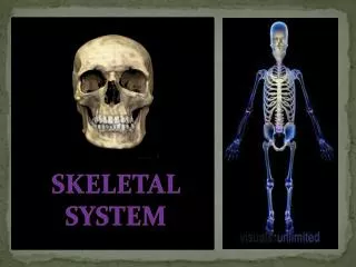

SKELETAL SYSTEM
E N D
Presentation Transcript
Skeletal system is the system of bones, associated cartilages and joints of human body. Together these structures form the human skeleton. Skeleton can be defined as the hard framework of human body around which the entire body is built.
TYPES OF BONES AXIAL BONES APPENDICULAR BONES
WHAT IS AXIAL BONES? • The axial skeleton forms the central axis of the body. It consists of the skull, the vertebral column, the ribs and the sternum or breastbone.
SKULL The skull consists of 28 different bones (including the ossicles of the ear). The bones of the skull can be divided into two main groups: the cranium which encloses and protects the brain and the facial bones.
VERTEBRAL COLUMN • The vertebral column forms the central part of the skeleton. It supports the skull and protects the spinal cord. It also serves as attachment for the ribs, the pectoral and pelvic girdles. The vertebral column consists of separate bones, the vertebrae. The different vertebrae are arranged above each other. Because the separate vertebrae are attached to each other by means of fibrous cartilaginous discs they form a flexible column. Each vertebra has articular surfaces above and below, which allow articulation movement between them. • The vertebral column of 33 vertebrae is divided into five regions according to their position and structure. The five regions consist of: Seven cervical (neck) vertebrae, Twelve thoracic (chest) vertebrae, Five lumbar vertebrae, Five fused sacral vertebrae, and Four fused vertebrae.
TYPICAL VERTEBRA A typical vertebra consists of the centrum, a neural arch, a neural spine, two transverse processes and four articular processes with articulating surfaces. The centrum is the front part (anterior) and consists of a solid piece of spongy bone encircled.
CERVICAL VERTEBRA • The neck region consists of 7 cervical vertebrae. These are the smallest vertebrae in the vertebral column. The first two cervical vertebrae are known as theatlas and axis. They are specially adapted to support the skull and to enable it to move. They differ from the structure of the typical vertebra in certain respects. • The Atlas • The Axis
CERVICAL VERTEBRA ATLAS AXIS
CERVICAL VERTEBRA (AXIS) • The axis has a large, strong neural spine. The centrum is small and has become modified to bear the odontoid process (a tooth-like projection) on its upper surface. The odontoid process fits against the facet in the anterior arch of the atlas. This forms a pivot joint or axis, around which the atlas (together with the skull) can rotate, so allowing the head to turn from side to side.
Cervical vertebrae (atlas) • The atlas is the first neck vertebra and supports the skull. It is ring-shaped and has no centrum. A neural spine is absent. The atlas consists ofposterior and anterior neural arches and 2 short transverse processes. The spinal foramen (neural canal) is very large. The 2 occipital condylesof the skull fit into the articulating facets on the upper surface of the atlas, on either side of the neural canal. On its lower surface (inferior) surface the atlas has2 articular surfaces for articulation with the axis.
THORACIC VERTEBRAE • It should be noted that all vertebrae are dorsal, although only 12 are thoracic. The 12 vertebrae of the thorax bear the ribs. The T1 vertebra (like C.V.7) is transitional in appearance. T2 to 8 vertebra are typical thoracic vertebrae with a kidney shaped body (fig. 39-5).
PARTS OF SKELETAL SYSTEM • Skull: • it occurs in the head with total 29 bones. Skull has four parts – cranium, face, bones of middle ear and hyoid. • Cranium: 8 bones • Face: 14 bones • Bones of middle ear: 6 bones • Hyoid: 1 bone
RIBS they are 12 pairs of bony bars that extend from thoracic vertebrae towards the sternum. First seven pairs are true ribs as they are attached to sternum directly. Next three are vertebrochondral or false ribs as they are attached to costal cartilage of seventh. 11thth pairs of ribs are imperfectly formed and do not reach the sternum and hence called floating ribs. and 12
STERNUM Sternum or breast-bone: it is a flat, narrow but about 15 cm long dagger-like bone present in the middle front part.
PECTURAL GIRDLE it consists of two halves, each with a clavicle and a scapula. Clavicle is twice-curved f-shaped thin elongated bone that extends from acromium process of scapula to manubrium part of sternum. Scapula is a thin curved triangular bone present on backside of thorax over second to seventh rib.
ARMS BONES each upper limb has 30 bones - along humerus in upper arm, two long curved radius and ulna in fore-arm, 8 carpals in wrist, 5 metacarpals in palm and 14 phalanges in fingers.
PELVIC GIRDLE it is a trough like bony structure formed by union of two similar halves or hip bones with themselves anterior and with the sacrum posteriorly. Each innominate is formed by union of 3 bones – ischium, pubis and ilium.
LEG BONES each lower limb has 30 bones – 1 femur, 1 patella, 1 tibia, 1 fibula, 7 tarsals, 5 metatarsals and 14 phalanges
CONCLUSION ON PARTS AND FUNCTION OF SKELETAL SYSTEM • Movements in vertebrates are performed by skeletal system and muscular system. Locomotion and movement of most external body parts are brought about by coordination of both the systems.
IMPOTANCE OF SKELETAL SYSTEM IT SERVE AS A FRAMEWORK IN OUR BODY TO PROTECT OUR BODY FROM SOME HARD 5THINGS AND GIVES US A BALANCE
SOURCES,,,,,,,,,,,,,,,,,,,, • Joel DeLisa and Walter C. Stolov, "Significant Body Systems," in: Handbook of Severe Disability, edited by Walter C. Stolov and Michael R. Clowers. US Department of Education, Rehabilitation Services Administration, 1981, pages 19-30. • Catherine Parker Anthony and Gary A. Thibodeau, Textbook of Anatomy & Physiology. St. Louis: Mosby, 1983, pages 328-346. • Anatomy Clipart (Designs4Free) • Dennis Kunkel, Electron Microscopy Gallery • J. Crimando, Anatomy and Physiology Tutorials (Maricopa)
SUBMITTED BY: CARLA CABALTICAN VERONICA, SY
SUBMITTED TO: SIR DELLOSA • PROJECT • IN • BIOLOGY 1-S